Latin: saccus lacrimalis
Overview
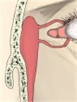
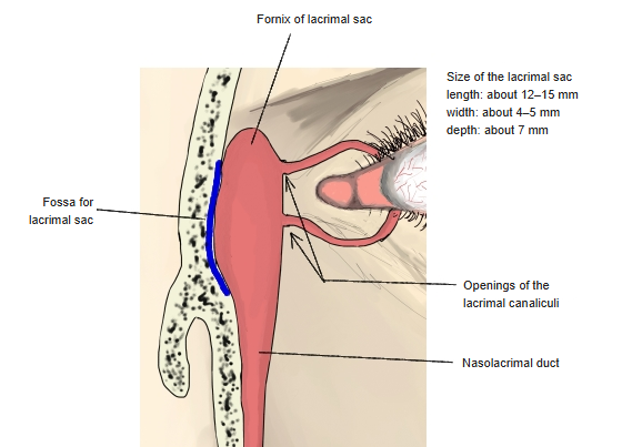
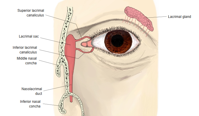
- The upper, widened end of the tear drainage passage, near the root of the nose.
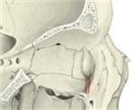
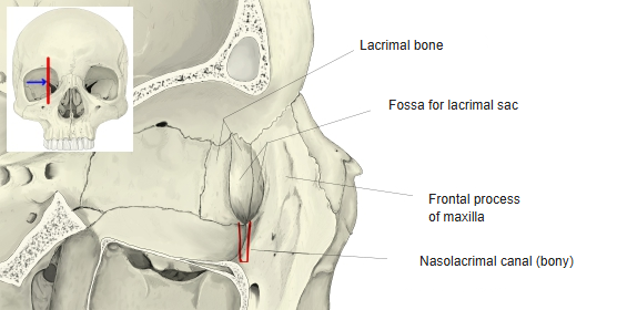
- Lies in the fossa for the lacrimal sac, formed by the lacrimal bone and the frontal process of the maxilla, behind the medial palpebral ligament.
- About 12–15 mm long and 4–8 mm wide; its closed upper end is the fornix of the lacrimal sac.
- The upper and lower lacrimal canaliculi open into its lateral wall; below it continues into the nasolacrimal duct.
Lacrimal sac 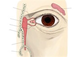
Lacrimal apparatus
Wall
- Lined by a tall columnar epithelium, partly ciliated, with goblet cells; the connective tissue under it contains many lymphocytes.
- Its mucosa is only loosely attached to the periosteum of the fossa.
- Contraction of the lacrimal part of orbicularis oculi during blinking helps to pump tears into the sac.
Bony nasolacrimal canal
References and sources
- Further reading: Lacrimal sac, Wikipedia.