Latin: ductus nasolacrimalis
Overview
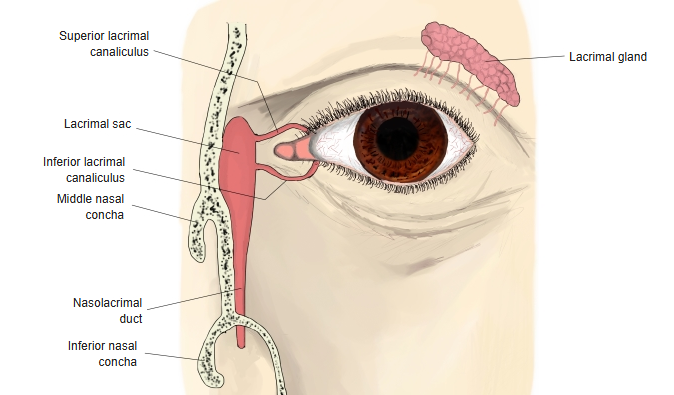
- The duct that carries tears from the lacrimal sac to the nose.
- About 12–24 mm long; runs downward and slightly backward and laterally.
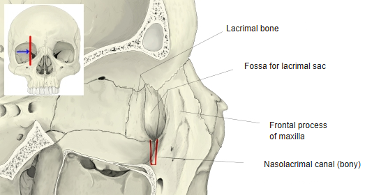
- A membranous duct inside the bony nasolacrimal canal, formed by the maxilla, the lacrimal bone and the inferior nasal concha.
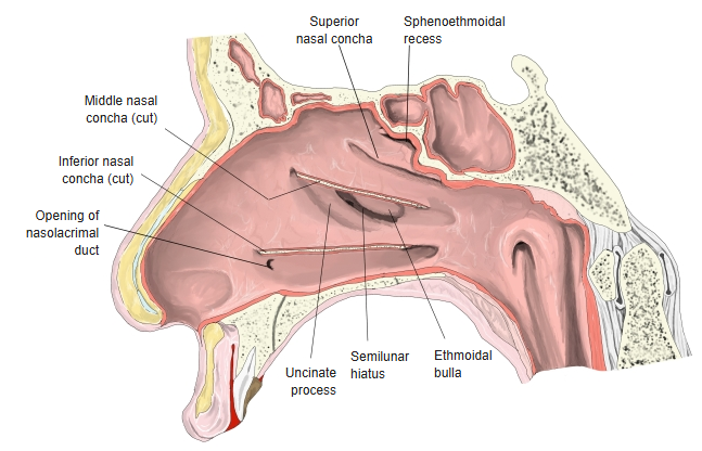
- Opens into the inferior nasal meatus, under the inferior nasal concha, about 30–35 mm behind the nostril.
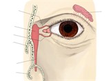
Lacrimal apparatus 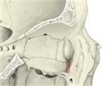
Bony nasolacrimal canal
Opening and lacrimal fold
- The shape of the opening varies: a wide round opening when it lies at the level of the bony canal, a vertical slit when it lies lower.
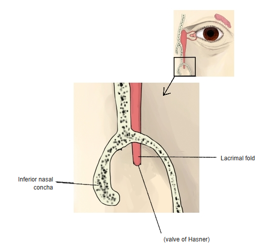
- A fold of mucosa over the opening, the lacrimal fold (valve of Hasner), helps to stop air and mucus being forced up the duct.
Opening of the nasolacrimal duct 
Lacrimal fold
Wall
- The mucosa is like that of the lacrimal sac: tall, partly ciliated columnar epithelium with goblet cells; mucous glands occur in the lower part.
- A dense venous plexus, continuous with that of the inferior nasal concha, lies between the mucosa and the periosteum – its swelling in a cold can block the duct and make the eyes water.
References and sources
- Further reading: Nasolacrimal duct, Wikipedia.