Latin: pleura
Overview
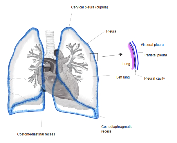
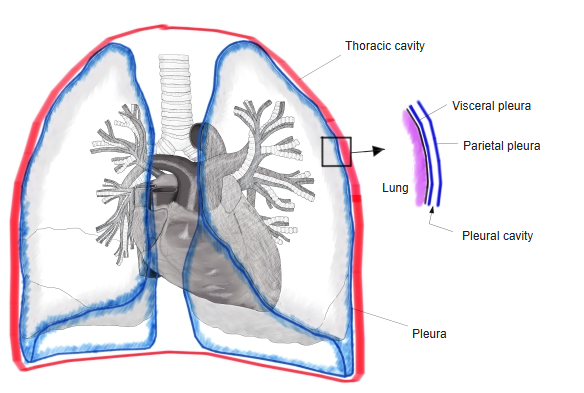
- A thin, transparent serous membrane that covers each lung and lines the walls of the thoracic cavity around it.
- The two layers enclose the pleural cavity, a narrow space holding a thin film of serous fluid; the right and left cavities are separated by the mediastinum.
- The fluid lets the lung surface slide freely against the chest wall during breathing.
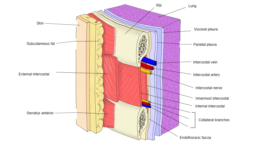
- Its surface is a single layer of mesothelium on a thin layer of connective tissue; outside the parietal pleura lies the endothoracic fascia.
Pleura and pleural recesses 
Pleural cavity
Parts
| Part | Covers |
|---|---|
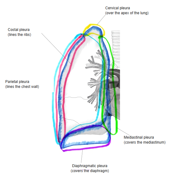
| Visceral (pulmonary) pleura | The surface of the lung, dipping into the fissures |
| Parietal pleura – costal part | The inner surface of the ribs and intercostal spaces |
| Parietal pleura – mediastinal part | The side of the mediastinum |
| Parietal pleura – diaphragmatic part | The upper surface of the diaphragm |
| Cervical pleura (cupula) | The dome over the apex of the lung; strengthened by thickened endothoracic fascia (suprapleural membrane) |
- At the hilum the visceral pleura turns back into the mediastinal pleura; below the root the two layers form the pulmonary ligament.
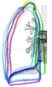
Parts of the parietal pleura 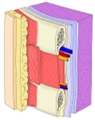
Layers of the chest wall
Pleural recesses
- Narrow parts of the pleural cavity where the parietal pleura folds from one wall to another; the lung enters them only in deep inspiration.
- Costodiaphragmatic recess: where the costal pleura meets the diaphragmatic pleura, low at the side and back – the lowest part of the cavity, where fluid collects.
- Costomediastinal recess: where the costal pleura meets the mediastinal pleura behind the sternum; larger on the left, at the cardiac notch.
References and sources
- Further reading: Pleura, Wikipedia.