Lung (Latin: pulmo)
- Overview
- External features (apex, base, surfaces, borders)
- Hilum and root of the lung
- Lobes
- Position
- Branching of the bronchi
- Structure
- Vessels and nerves
- Development
- Related terms
- References
Overview
system
- Airway
- Nasal cavity : from the nostrils to the choanae; divided into right and left halves by the nasal septum and lined with mucosa.
- Pharynx : made of three parts (upper, middle, lower) and lined with mucosa; also part of the digestive system.
- Larynx : funnel-shaped tube at the level of the 4th–7th cervical vertebrae; its framework is cartilage.
- Trachea : tube about 10 cm long and 20–25 mm wide; divides at the level of the 4th–5th thoracic vertebrae.
- Bronchi : branch from the trachea to the right and left, and divide into many bronchial branches inside the lungs.
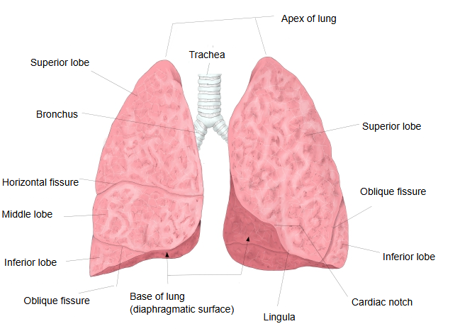
- Lungs : right lung (upper, middle, lower lobes), left lung (upper, lower lobes); gas exchange (external respiration) takes place in the alveoli.
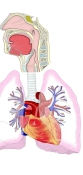
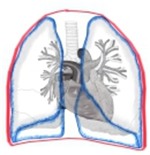 Thorax
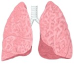 Lungs (anterior view)
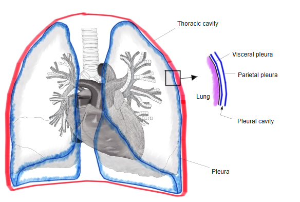
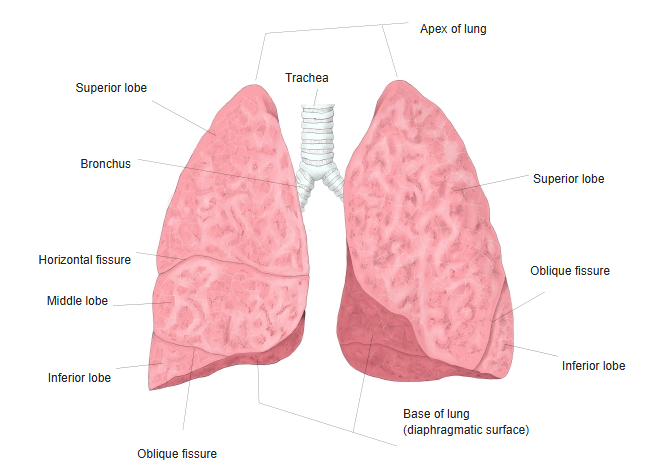
In humans the two lungs together weigh about 1 kg in men and about 0.9 kg in women. They occupy most of the thoracic cavity and consist mainly of airways and blood vessels, which meet in the alveoli for gas exchange. There are about 300 million alveoli, with a total surface area of about 70 m².
External features
· The colour is usually pearly grey to pink.
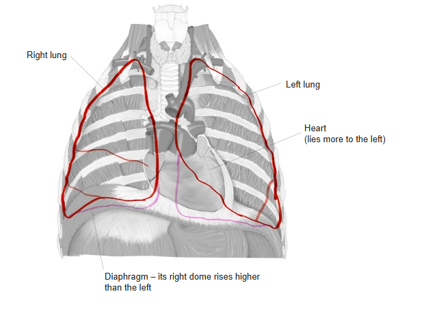
· The left lung lies to the left of the trachea, esophagus, heart and aorta; the right lung lies to the right of them.
· The left lung is narrower and longer than the right, smaller in volume (left 8 : right 10), and reaches a little lower.
· Together the lungs weigh about 1 kg in men and about 0.9 kg in women; the right lung (about 1200 mL, 600 g) is a little larger than the left (about 1100 mL, 500 g).
The two lungs differ in shape because:
· the heart lies more to the left;
· the right lobe of the liver is large;
· the right half of the diaphragm bulges higher than the left half.
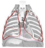 Left and right lungs
Each lung has an upper end (apex), a lower base, and surfaces and borders.
- Apex : the blunt, rounded top that rises 2–3 cm above the clavicle and is covered by the cervical pleura.
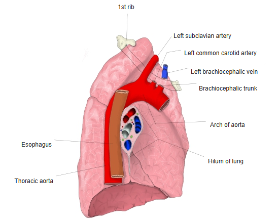
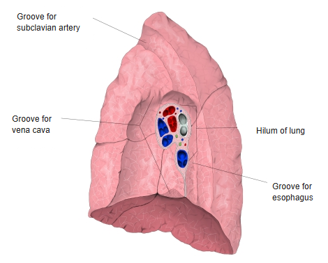
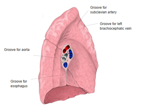
Groove for the subclavian artery (both lungs), groove for the left brachiocephalic vein (left), groove for the superior vena cava (right). - 3 surfaces : costal surface (the largest, facing the ribs), mediastinal (medial) surface (where the bronchi, vessels and nerves enter and leave), diaphragmatic surface (= the base).
- 3 borders : anterior border (between the costal and medial surfaces in front), inferior border (between the costal and diaphragmatic surfaces), posterior border (between the costal and medial surfaces behind).
- Base : hollowed to fit the dome of the diaphragm; = the diaphragmatic surface.
[Apex of the lung]
· It is covered by the cervical pleura.
On the medial side of both apices the subclavian artery makes the groove for the subclavian artery. In addition, the left lung has a groove for the left brachiocephalic vein, and the right lung has a groove for the superior vena cava.
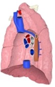 Right lung
(medial surface)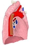 Left lung
(medial surface)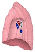 Grooves of the right lung
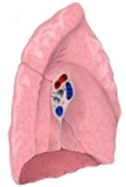 Grooves of the left lung
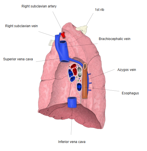
[Base of the lung]
[Surfaces]
Three surfaces are distinguished on each lung.
the lung
- Costal surface : the outer surface facing the ribs; the largest surface. It may show rib impressions (the 1st rib and sometimes others).
- Medial surface : faces the mediastinum; as a whole slightly hollowed, deeply so at the cardiac impression (the hollow for the heart). It contains the hilum.
- Mediastinal part : the front part facing the mediastinum (see the note below).
- Vertebral part : the back part facing the vertebral bodies.
- Diaphragmatic surface : the lower surface facing the diaphragm; the same as the base, hollowed to fit the dome of the diaphragm.
Note: some descriptions use "mediastinal part" in a narrower sense, for the medial surface excluding the vertebral part and the cardiac impression.
[Borders]
Three borders are distinguished; all three are edges between surfaces.
the lung
- Anterior border : the sharp edge between the costal surface and the front of the medial surface.
- Inferior border : the sharp edge between the costal and diaphragmatic surfaces; it is less sharp where it turns onto the medial surface.
- Posterior border : the blunt edge between the costal surface and the back of the medial surface.
- Left and right lungs
 Surfaces of the lung
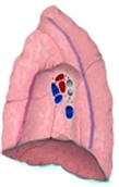 Borders
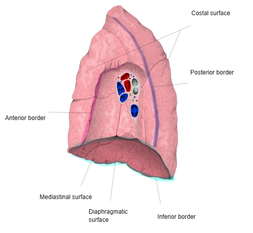
Hilum and root of the lung
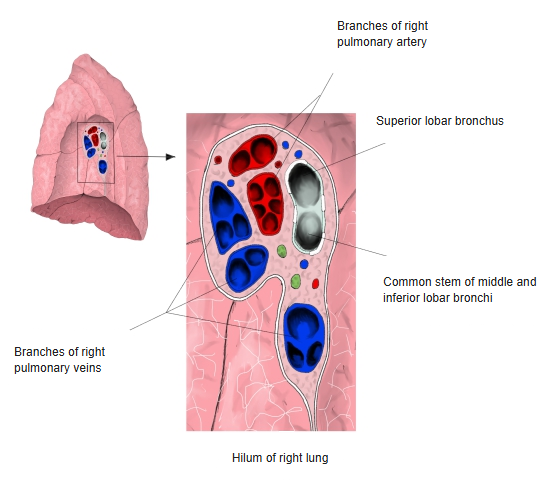
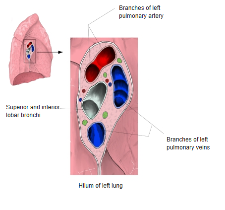
· At the hilum the bronchus lies furthest back, the pulmonary artery in front of it, and the pulmonary veins further in front.
· The hilum lies in the middle of the medial surface, at about the level of the 6th thoracic vertebra.
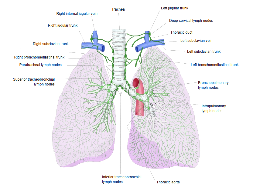
· Around the hilum the bronchopulmonary lymph nodes are well developed and collect the lymph from inside the lung.
At the hilum the pulmonary (visceral) pleura covering the lung wraps around the root of the lung and turns into the parietal pleura lining the wall of the thoracic cavity. The root is covered by pleura on its upper, front and back sides. Below the root the front and back layers come together and form a double fold that runs from the lower part of the hilum towards the diaphragmatic surface: the pulmonary ligament.
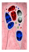 Hilum of the right lung
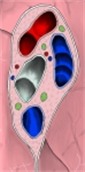 Hilum of the left lung
Lobes
Because the pulmonary pleura runs into the oblique and horizontal fissures, the lobes are almost completely separate and are joined only by their bronchial branches (and vessels).
 Lungs (anterior view)
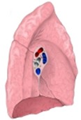 Left lung
(medial surface)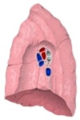 Right lung
(medial surface)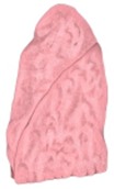 Left lung
(lateral surface)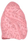 Right lung
(lateral surface)
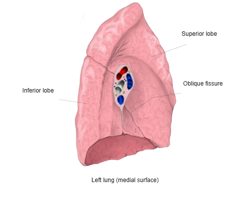
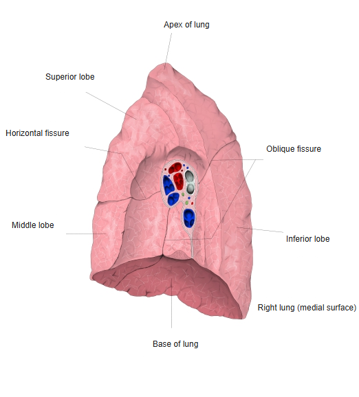
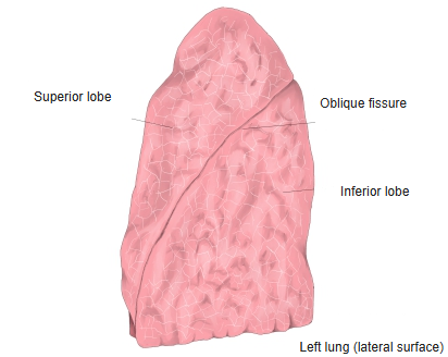
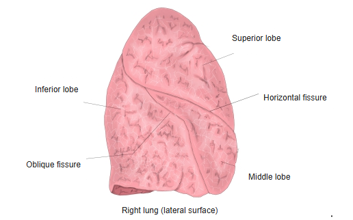
Position
· The left lung lies to the left of the trachea, esophagus, heart and aorta, and the right lung to the right of them.
[Anterior limit]
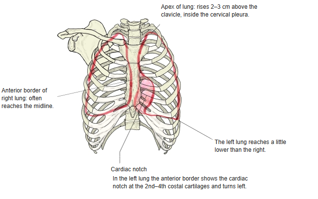
The anterior limit follows the anterior border of the lung. On both sides it starts behind the sternoclavicular joint, curves towards the other side as it descends, and at the level of the 6th costal cartilage turns into the lower limit. On the left, however, it swings away from the midline at the level of the 2nd–4th costal cartilages, forming the clear cardiac notch, while on the right it often reaches the midline.
· The left lung is narrower and longer than the right, smaller in volume (left 8 : right 10), and reaches a little lower.
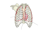 Thorax and lungs
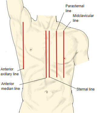
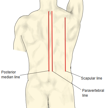
(anterior view)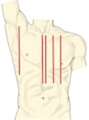 Vertical reference lines
(front)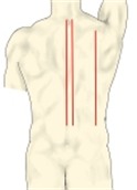 Vertical reference lines
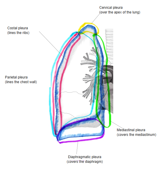
(back)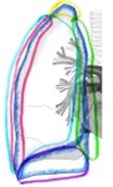 Parts of the pleura
Branching of the bronchi
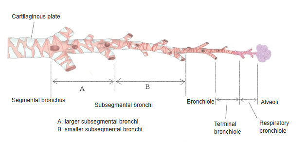
Inside the lungs the right and left bronchi branch as follows: lobar bronchi → segmental bronchi → bronchioles → terminal bronchioles → respiratory bronchioles → alveolar ducts → alveolar sacs → alveoli.
| 1 | Lobar bronchi | Branch from the main bronchi at the hilum (2 on the left, 3 on the right). Inside the lung their structure is almost the same as that of the main bronchi, with some differences: · They have no membranous wall. · The C-shaped cartilages gradually shrink into irregular plates (found down to branches about 1 mm wide). · The cells of the pseudostratified ciliated columnar epithelium gradually become lower. |
| 2 | Segmental bronchi | · Each lobar bronchus divides into 2–5 segmental bronchi. · The region supplied by the branches of one segmental bronchus is a bronchopulmonary segment; the segments have fixed numbers. · Irregular cartilage plates are present. |
| 3 | Bronchioles | · The segmental bronchi divide into many segmental bronchial branches. · At the end of these branches, the thin stem that enters a pulmonary lobule is a bronchiole (0.8–1 mm wide). · Within the lobule it divides into 3–6 smaller branches. The cone-shaped region, 1–2 cm across, supplied by one bronchiole is the pulmonary lobule. In the wider bronchioles the epithelium is pseudostratified ciliated epithelium with goblet cells, the lamina propria contains bronchial glands (mixed glands), smooth muscle runs beneath it, and below the muscle is a layer of loose connective tissue with cartilage plates. When a bronchiole becomes narrower than 1 mm, cartilage disappears and the smooth muscle becomes stronger and runs circularly. Excessive contraction of this muscle narrows the airways, as in bronchial asthma. |
| 4 | Terminal bronchioles | · Within the pulmonary lobule the bronchioles divide into terminal bronchioles. · Their epithelium is simple columnar, made of ciliated cells and secretory cells, with a thin layer of scattered smooth muscle beneath it. |
| 5 | Respiratory bronchioles | · Alveoli open from their walls here and there. · Each respiratory bronchiole gives rise to 2–9 alveolar ducts, which end in blind alveolar sacs surrounded by alveoli. · Their epithelium is simple columnar to simple cuboidal, resting on thin connective tissue with a little smooth muscle. |
 Segmental bronchus
to alveolar sacs
Structure
the lung
- Conducting part : main bronchi → lobar bronchi → segmental bronchi → bronchioles → terminal bronchioles.
- Respiratory part : respiratory bronchioles, alveolar ducts, alveolar sacs, alveoli.
Vessels and nerves
The vessels and nerves of the lungs are as follows.
of the lung
- Functional vessels : the vessels that carry blood for gas exchange in the alveoli: pulmonary arteries and pulmonary veins.
- Nutrient vessels : the vessels that supply the lung tissue itself: bronchial arteries and bronchial veins.
[Functional vessels]
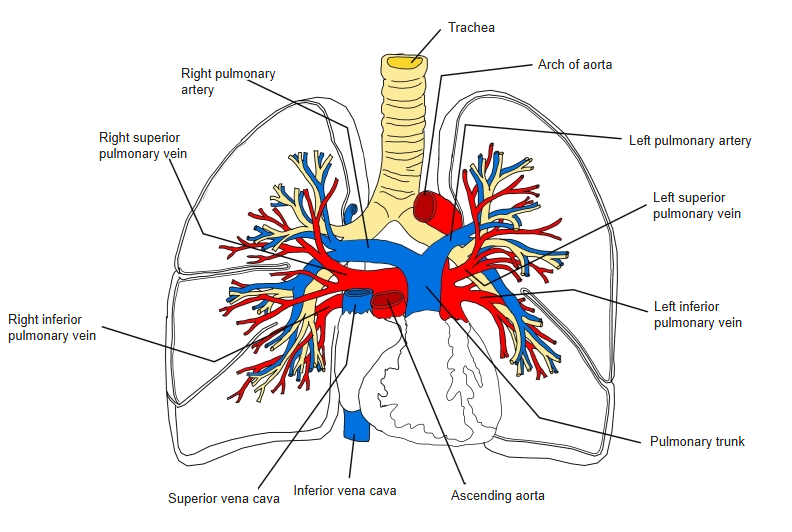
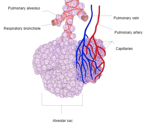
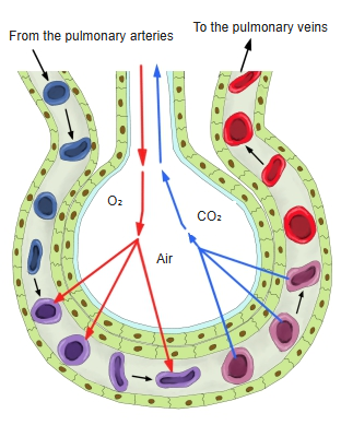
The pulmonary arteries, which bring venous blood to the lungs, are one on each side. Each enters at the hilum, branches alongside the bronchi, and surrounds the alveoli with a capillary network. Gas exchange takes place here, and the oxygen-rich blood enters the veins, which run along the bronchi, gradually join into larger trunks and finally leave the hilum as the pulmonary veins.
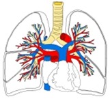 Pulmonary arteries
and veins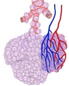 Alveolar sac with
arteries and veins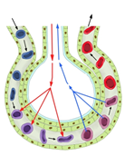 Gas exchange
[Nutrient vessels]
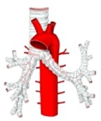
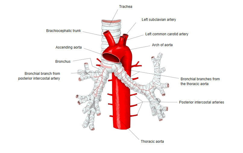
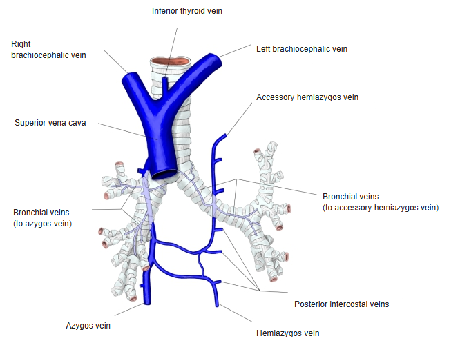
The bronchial arteries run with the branching bronchi. They arise from the thoracic aorta as 2–3 vessels or by a common trunk, divide to the right and left, and supply the muscle, glands and vessel walls of the bronchi and lung, and also the pleura. The blood from them returns through the bronchial veins, which leave the hilum and drain into the azygos vein on the right and into the accessory hemiazygos vein or the left superior intercostal vein on the left.
 Bronchial arteries
 Bronchial veins
[Lymphatics]
Lymph vessels on the surface of the lung form the subpleural lymphatic plexus under the pulmonary pleura and drain to the bronchial and pulmonary lymph nodes. Deeper vessels run around the alveoli, alveolar ducts and bronchi and drain to the bronchopulmonary lymph nodes at the hilum.
 Bronchi and lungs
[Nerves]
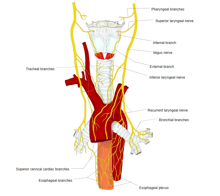
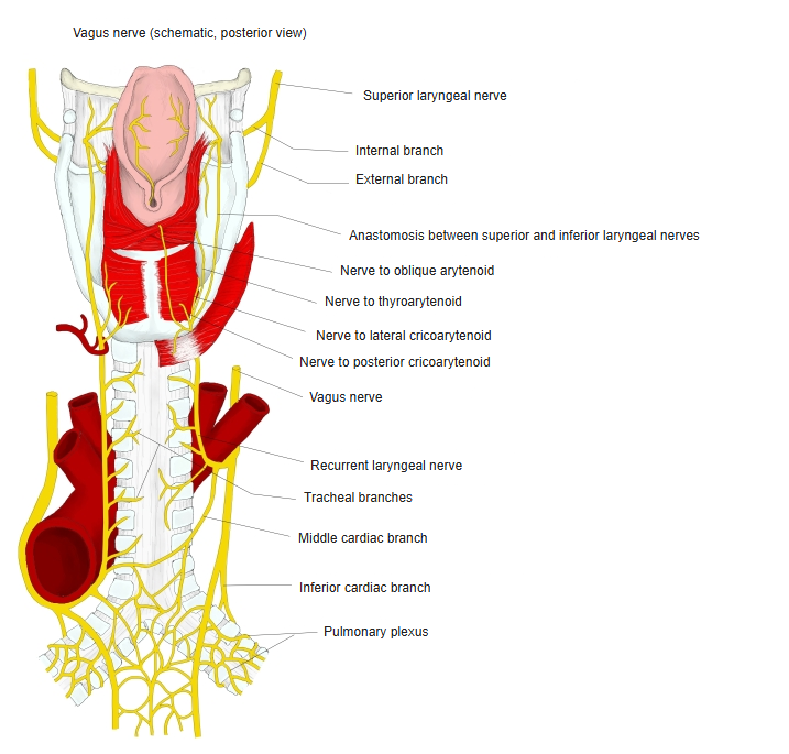
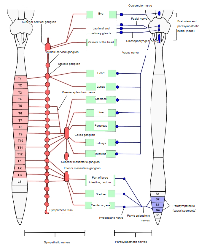
The lungs are supplied by the vagus nerve and by sympathetic nerves. From the hilum the nerves branch along the bronchi and form plexuses. The vagus nerve makes the bronchial muscle contract and the vessels dilate; the sympathetic nerves act against it. The vagus fibres enter the root of the lung as the anterior and posterior pulmonary plexuses, and sympathetic fibres from the inferior cervical (cervicothoracic) ganglion and the cardiac ganglia also enter here. Small groups of sympathetic nerve cells may be found along the branching bronchi.
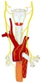 Nerves 1
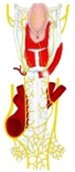 Nerves 2
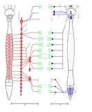 Autonomic nerves
Development
The respiratory primordium arises as a blind pouch on the front wall of the foregut. At the end of the 4th week of embryonic life its end divides to form the bronchi; the right side divides into three and the left into two, matching the number of lobes in the finished lungs. Repeated further branching follows, and the ends of the branches enlarge to form the bronchioles, terminal bronchioles and primitive alveoli, which become the alveolar ducts. Late in fetal life alveoli develop especially rapidly, budding from the respiratory bronchioles, and new alveoli continue to form in early childhood.
The epithelium of the developing alveoli is ciliated until late in fetal life; it then loses its cilia, and when air enters after birth the alveolar ducts widen and the epithelium of the alveolar walls becomes flat. The connective tissue, elastic fibres and cartilage of the lung arise from the mesoderm.
Related terms
Main anatomical terms related to the lung, in alphabetical order.
References and sources
The two lungs lie in the thoracic cavity on either side of the heart and the mediastinum. Their task is gas exchange: oxygen passes from the air into the blood, and carbon dioxide from the blood into the air. The thin, moist pleurae reduce friction between the lungs and the chest wall during breathing, and the main muscle that drives breathing is the diaphragm.
The right lung is larger than the left, which shares the left side of the chest with the heart. The airways that receive the inhaled air form the conducting zone, which ends at the terminal bronchioles; the respiratory zone begins with the respiratory bronchioles, which lead into alveolar ducts, alveolar sacs and alveoli, where gas exchange takes place. Each lung lies in a pleural sac whose two layers are separated by a thin film of pleural fluid. The lungs have two blood supplies: the pulmonary circulation, which brings deoxygenated blood from the heart for gas exchange, and the bronchial circulation, which brings oxygenated blood to the lung tissue itself.
In the embryo the lungs develop as an outgrowth of the foregut. Before birth they are filled with fluid and blood largely bypasses them through the ductus arteriosus; at birth air enters the lungs, the ductus closes, and the lungs begin to work. They continue to develop in early childhood.
- Further reading: Lung, Wikipedia.