Latin: glandula thyroidea
Overview
- The largest endocrine gland, in the front of the neck below the larynx.
- Two lobes joined by an isthmus in front of the 2nd–4th tracheal rings; weighs about 20–25 g.
- Makes the thyroid hormones T4 and T3 (metabolism) and calcitonin.
- Enclosed in pretracheal fascia, so it moves up and down when swallowing.
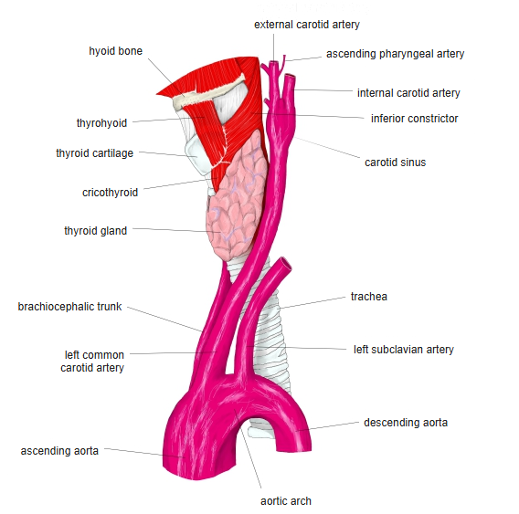
Thyroid gland and the great arteries
Relations and structure
- Each lobe extends from the side of the thyroid cartilage to about the 6th tracheal ring.
- Covered by the infrahyoid muscles; the common carotid artery lies behind and lateral to each lobe.
- The parathyroid glands lie on the back of the lobes; the recurrent laryngeal nerve runs close behind.
- A pyramidal lobe may rise from the isthmus (remnant of the thyroglossal duct).
- Microstructure: follicles lined by follicular cells, filled with colloid (thyroglobulin); parafollicular C cells secrete calcitonin.
Blood supply
| Artery | Origin |
|---|---|
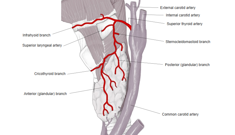
| Superior thyroid artery | External carotid artery |
| Inferior thyroid artery | Thyrocervical trunk of the subclavian artery |
| Thyroid ima artery (inconstant) | Brachiocephalic trunk or aortic arch |
- Veins: superior and middle thyroid veins to the internal jugular vein; inferior thyroid veins to the brachiocephalic veins.
Superior thyroid artery
References and sources
- Further reading: Thyroid gland, Wikipedia.