Medial surface of fibula (Latin: facies medialis fibulae)
Also called the anterior surface or the extensor surface
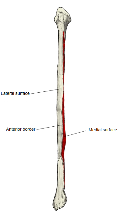
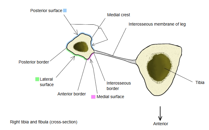
One of the three surfaces of the shaft of the fibula: the narrow surface between the anterior border and the interosseous border.
- Medial surface : a narrow surface between the anterior border and the interosseous border, facing forwards and medially.
- Lateral surface : a fairly flat surface between the anterior border and the posterior border.
- Posterior surface : the surface between the posterior border and the interosseous border; it bears the medial crest.
It is very narrow in its upper third and becomes wider in its lower third, where it may be slightly hollowed into a shallow groove.
This slightly hollowed, narrow surface lies between the interosseous membrane behind and the anterior intermuscular septum in front, and gives origin to the extensor muscles of the front of the leg.
Note on names: many older English texts (for example Gray's Anatomy) call this surface the anterior surface (extensor surface) and use "medial surface" for the part of the posterior surface in front of the medial crest, from which tibialis posterior arises. This page follows Terminologia Anatomica.
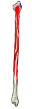 Muscle attachments (posterior)
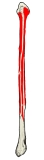 Muscle attachments (anterior)
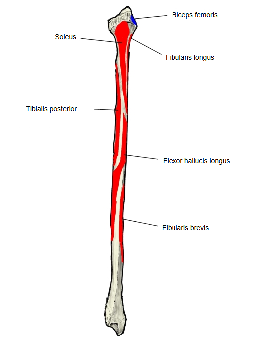
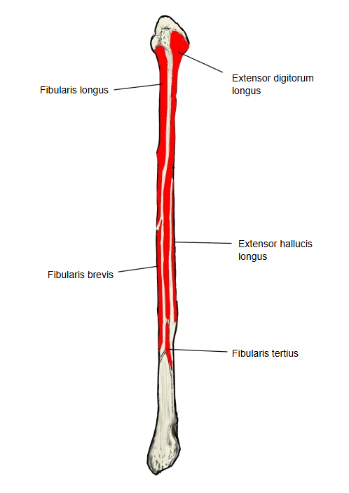
Attachments
| Muscles | 1 | Extensor digitorum longus | upper part of the surface |
| 2 | Extensor hallucis longus | middle part | |
| 3 | Fibularis tertius (peroneus tertius) | lower part |
Where it is found
Related terms
- Shaft of fibula
- Anterior border of fibula
- Interosseous border of fibula
- Lateral surface of fibula
- Posterior surface of fibula
- Medial crest of fibula
References and sources
- Further reading: Fibula, Wikipedia.
- Federative International Programme for Anatomical Terminology (FIPAT). Terminologia Anatomica, 2nd ed. (TA2), 2019.