Shaft of fibula (Latin: corpus fibulae)
Also called the body of fibula
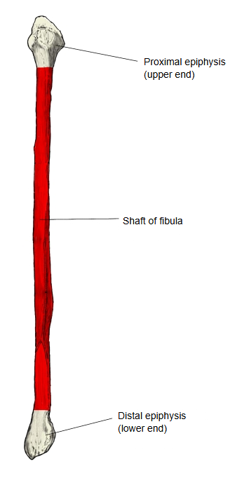
The shaft (body) of the fibula is the long middle part of the fibula when the bone is divided into three parts; it is the diaphysis, and three borders and three surfaces can be distinguished on it.
- Proximal end (upper end) : the swollen head of fibula; it gives attachment to muscles and ligaments and has a joint surface for the tibia
- Shaft of fibula : the diaphysis; a three-sided prism with 3 borders and 3 surfaces
- Distal end (lower end) : the thick lateral malleolus; it articulates with the talus and is bound to the tibia by ligaments
Anatomy textbooks describe the shaft as follows.
The shaft is a long, slender three-sided prism and is slightly twisted. Its anterior, posterior and interosseous borders divide it into three surfaces. Because the shaft is buried in the leg muscles, its surfaces are where those muscles arise, and its borders give attachment to the connective-tissue sheets that separate the muscles or muscle groups of the leg: the intermuscular septa and the interosseous membrane. For this reason the sharpness of the borders, the width of the surfaces and the amount of twist vary a good deal between people, with the development of the muscles.
Question: does the neck of fibula belong to the proximal end or to the shaft? It is usually described on its own, as the narrow part just below the head that joins the head to the shaft, so it can be seen as belonging to both.
Three borders
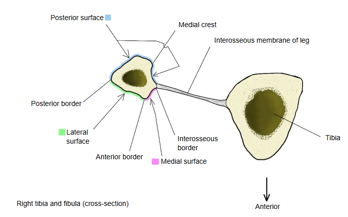
The shaft of the fibula has the following three borders.
- Anterior border : the sharpest of the three; it starts in front of the head and divides near the lower end to enclose a triangular area above the lateral malleolus
- Posterior border : blunt and rounded, especially above; it runs from the apex of the head to the back of the lateral malleolus
- Interosseous border : a fairly sharp ridge facing the tibia, for the interosseous membrane; its upper part is often indistinct
In addition to these three borders, the fibula has a ridge called the medial crest, which runs down the posterior surface. The Wikipedia article on the fibula (from Gray's Anatomy) counts this crest as a border and so describes four borders.
Wikipedia describes four borders of the body of the fibula: antero-lateral, antero-medial, postero-lateral and postero-medial.
- Antero-lateral border : begins above, in front of the head ⇒ anterior border
- Antero-medial border : also called the interosseous crest ⇒ interosseous border
- Postero-lateral border : begins above at the apex of the head and ends below at the back of the lateral malleolus ⇒ posterior border
- Postero-medial border : begins at the medial side of the head ⇒ medial crest
Three surfaces
- Medial surface : a narrow surface between the anterior and interosseous borders, facing medially (and forwards)
- Lateral surface : a fairly flat surface between the anterior and posterior borders, facing outwards
- Posterior surface : the surface between the posterior and interosseous borders, the widest; the medial crest runs on it
The same Wikipedia article describes the body of the fibula as having four surfaces.
These are the anterior, posterior, medial and lateral surfaces. In this older description the medial crest divides the present posterior surface into a "posterior" and a "medial" surface, and the present medial surface is called the "anterior" surface.
- Anterior surface : between the antero-lateral and antero-medial borders
- Posterior surface : between the postero-lateral and postero-medial borders
- Medial surface : between the antero-medial and postero-medial borders
- Lateral surface : between the antero-lateral and postero-lateral borders
Attachments
| Muscles | 1 | Tibialis posterior (origin) | musculus tibialis posterior | 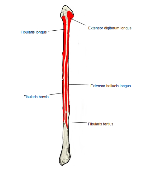 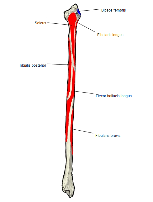 |
| 2 | Fibularis tertius (origin) | musculus fibularis tertius | ||
| 3 | Extensor digitorum longus (origin) | musculus extensor digitorum longus | ||
| 4 | Fibularis longus (origin) | musculus fibularis longus | ||
| 5 | Flexor hallucis longus (origin) | musculus flexor hallucis longus | ||
| 6 | Extensor hallucis longus (origin) | musculus extensor hallucis longus | ||
| 7 | Fibularis brevis (origin) | musculus fibularis brevis | ||
| 8 | Soleus (origin) | musculus soleus |
By surface: extensor digitorum longus, extensor hallucis longus and fibularis tertius arise from the medial surface; fibularis longus and fibularis brevis from the lateral surface; soleus, tibialis posterior and flexor hallucis longus from the posterior surface. Biceps femoris inserts on the head, not on the shaft.
Where it is found
Related terms
- Anterior border of fibula
- Posterior border of fibula
- Interosseous border of fibula
- Medial surface of fibula
- Lateral surface of fibula
- Posterior surface of fibula
- Medial crest of fibula
References and sources
- Further reading: Fibula, Wikipedia.
- Further reading: Interosseous membrane of leg, Wikipedia.
- Federative International Programme for Anatomical Terminology (FIPAT). Terminologia Anatomica, 2nd ed. (TA2), 2019.