Mandibular canal (Latin: canalis mandibulae)
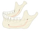 Mandibular canal (oblique view)
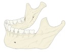 Mandible (lateral)
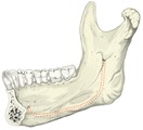 Mandibular canal (medial view)
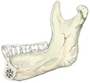 Mandible (medial surface)
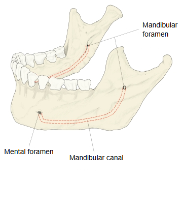
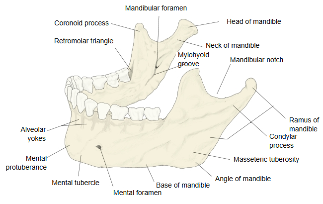
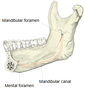
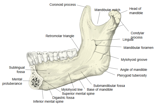
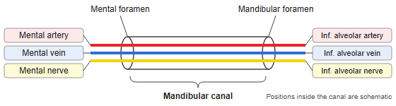
Inferior alveolar nerve: one of the terminal branches of the mandibular nerve (the third division of the trigeminal nerve). At the mental foramen it continues as the mental nerve.
Inferior alveolar artery: one of the branches of the first (mandibular) part of the maxillary artery. At the mental foramen it gives off the mental artery.
Inferior alveolar vein: the continuation of the mental vein after it enters the mental foramen; it runs back through the canal, leaves at the mandibular foramen and drains into the pterygoid venous plexus.
The mandibular canal carries the inferior alveolar artery, vein and nerve. It begins at the mandibular foramen on the ramus, runs downwards and forwards in the ramus and then forwards in the body of the mandible along its long axis, below the roots of the teeth, and opens at the mental foramen in the chin region. On its way it sends small canals to every tooth socket, for the vessels and nerves of the lower teeth and the gums. Below the premolars it divides: one branch turns up and back to the mental foramen, and a smaller incisive canal continues forwards to the canine and incisor teeth.
The canal lies close to the roots of the lower molars, especially the second and third molars; care is needed in root canal treatment, extractions and implants so as not to injure the inferior alveolar nerve.
Reference websites
[Wikipedia]
· Mandibular canal, Wikipedia.
· Inferior alveolar nerve, Wikipedia.
[Websites with illustrations]
· The mandible (TeachMeAnatomy)
· The mandible: anatomy, structure, function (Kenhub)
Where it is found
Related terms
References and sources
- Further reading: Mandibular canal, Wikipedia.
- Further reading: Inferior alveolar nerve, Wikipedia.
- Federative International Programme for Anatomical Terminology (FIPAT). Terminologia Anatomica, 2nd ed. (TA2), 2019.