Mylohyoid line (Latin: linea mylohyoidea)
Back end
Sources differ considerably on where the back end of the line lies.
Short version: it ends at the front of the inner surface of the ramus, below the last molar.
Long version: it continues up to the lingula of the mandible. Its back part then runs close to and roughly parallel with the mylohyoid groove.
Most illustrations found by an internet image search show the short version.
Mylohyoid: one of the suprahyoid muscles; it arises from almost the whole length of the mylohyoid line.
Superior pharyngeal constrictor: one of the pharyngeal constrictors; a small part of it arises from the back end of the line, near the alveolar margin.
Pterygomandibular raphe: a tendinous band of connective tissue that gives origin to the superior pharyngeal constrictor; its lower end is attached near the back end of the mylohyoid line.
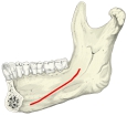 Mylohyoid line (medial view)
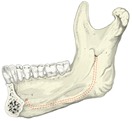 Right half of mandible (medial view)
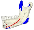 Mandible: muscle attachments (medial surface)
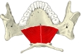 Mylohyoid (superior view)
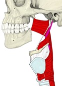 Pharyngeal constrictors (lateral view)
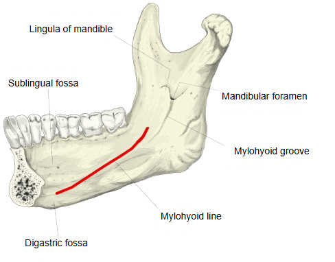
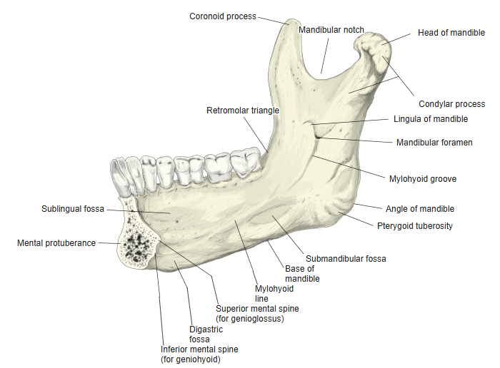
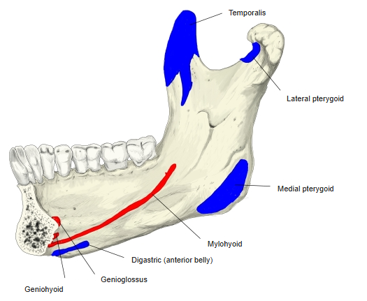
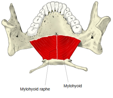
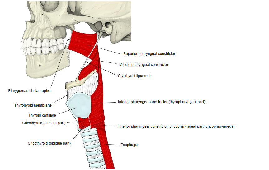
The line begins above and lateral to the digastric fossa and runs obliquely upwards and backwards, passing below the back end of the alveolar part. The mylohyoid arises from most of it and forms the muscular floor of the mouth; a small part of the superior pharyngeal constrictor arises from its back part. Below the line and parallel with it, the mylohyoid groove runs from the mandibular foramen on the inner surface of the ramus; it carries the mylohyoid nerve and vessels and is most distinct near the foramen. The line separates the sublingual fossa, above its front part, from the submandibular fossa, below its back part.
Reference websites
[Wikipedia]
· Mylohyoid line, Wikipedia.
· Mylohyoid muscle, Wikipedia.
[Websites with illustrations]
· The mandible (TeachMeAnatomy)
· Mandible, inner surface (Gray's Anatomy) (Wikimedia Commons)

Where it is found
Related terms
References and sources
- Further reading: Mylohyoid line, Wikipedia.
- Further reading: Mylohyoid muscle, Wikipedia.
- Federative International Programme for Anatomical Terminology (FIPAT). Terminologia Anatomica, 2nd ed. (TA2), 2019.