Latin: Arteria renalis
Overview
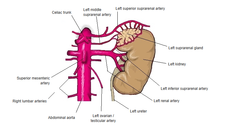
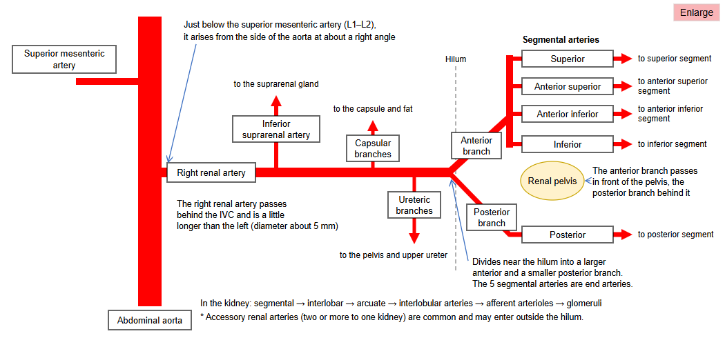
- A paired branch of the abdominal aorta arising at about the level of L1–L2, just below the superior mesenteric artery.
- The right renal artery is longer and passes behind the inferior vena cava; the left is shorter.
- Near the hilum of the kidney it divides into anterior and posterior branches, which give the segmental arteries.
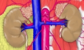
Renal artery 
Branches of the abdominal aorta
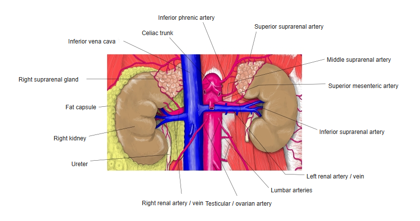
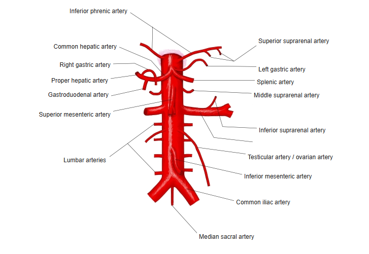
Branches
- Inferior suprarenal artery to the suprarenal gland.
- Ureteric branches to the upper ureter and capsular branches to the renal capsule.
- Segmental arteries (superior, anterior superior, anterior inferior, inferior and posterior) – each supplies one segment of the kidney with no anastomoses between them.
- Inside the kidney: interlobar → arcuate → interlobular arteries → afferent arterioles of the glomeruli.
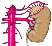
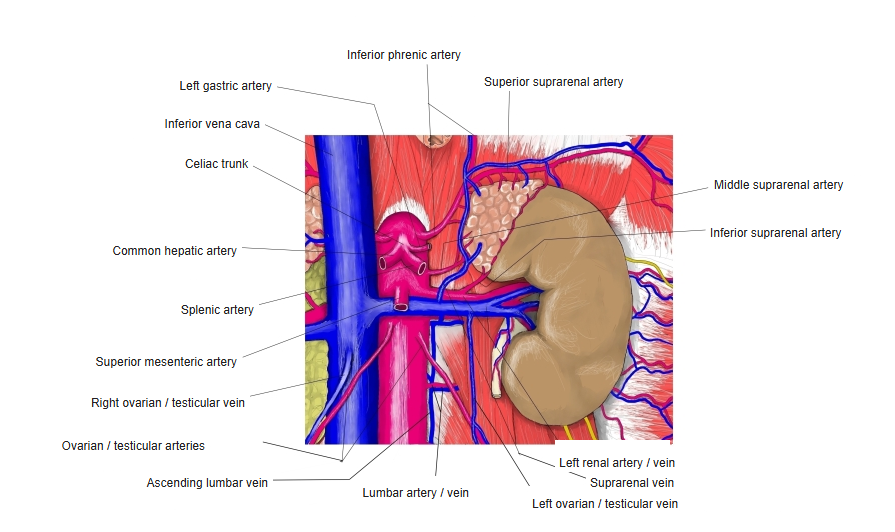
Arteries of the suprarenal gland and kidney 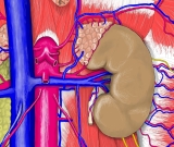
Celiac trunk and the vessels of the kidney 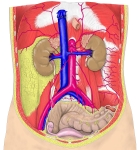
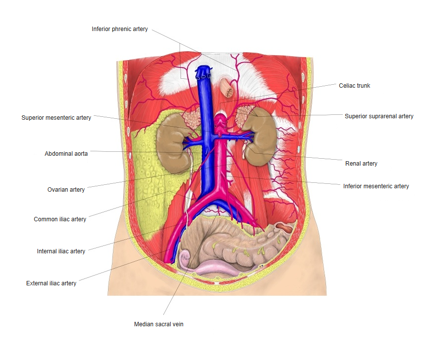
Abdominal aorta and its branches (front view) 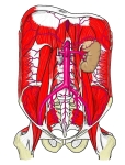
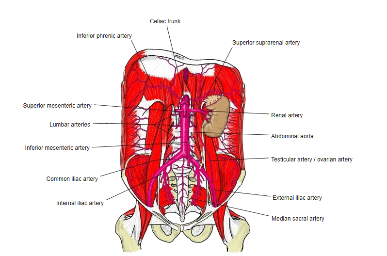
Abdominal aorta on the posterior abdominal wall
Clinical notes
- Accessory renal arteries are common.
- Narrowing of the renal artery (renal artery stenosis) can cause high blood pressure.
References and sources
- Further reading: Renal artery, Wikipedia.