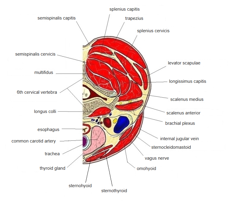
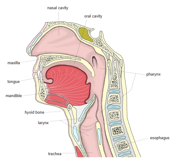
Transverse, sagittal and coronal sections of the neck and larynx. Select a figure to open a larger version with labels.
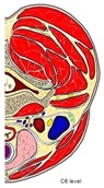
Transverse section of the neck at C6 (one side) - Back of the neck: trapezius, splenius capitis and semispinalis capitis around the cervical vertebra.
- Carotid sheath with the common carotid artery, internal jugular vein and vagus nerve.
- Front of the neck: sternocleidomastoid, thyroid gland and the airway near the midline.
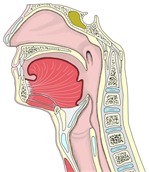
Midsagittal section from the nasal cavity to the larynx - Nasal cavity, oral cavity, pharynx and larynx in one view.
- Epiglottis, hyoid bone and laryngeal cartilages in front of the cervical vertebrae.
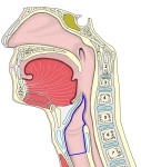
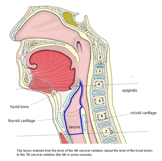
Larynx in a midsagittal section (small overview) - The larynx (blue outline) below the pharynx and in front of the oesophagus.
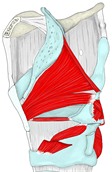
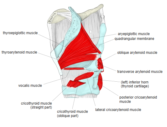
Intrinsic laryngeal muscles (median section of the larynx) - Intrinsic muscles of the larynx (red), such as the thyroarytenoid and cricoarytenoid muscles.
- Epiglottis and the thyroid, arytenoid and cricoid cartilages (pale blue).
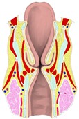
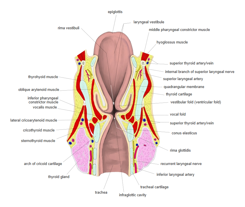
Coronal section of the larynx - Vestibular folds above and vocal folds below, with the laryngeal ventricle between them.
- Thyroid and cricoid cartilages (pale blue), infrahyoid muscles outside them and the thyroid gland (pink) at the bottom.
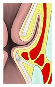
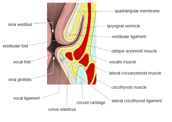
Vestibular fold and vocal fold (coronal section, enlarged) - Upper fold: the vestibular fold with the vestibular ligament.
- Lower fold: the vocal fold with the vocal ligament and the vocalis muscle.
Back to Cross-sections (all regions)
Other regions: Oral cavity, tongue and pharynx · Thorax and abdomen · Pelvis and perineum · Upper limb · Lower limb