Latin: auricula
Overview
- The visible part of the external ear (also called the pinna), on the side of the head.
- A thin plate of elastic cartilage covered by skin; the lower lobule contains only fat and connective tissue.
- Its folds funnel sound into the external acoustic meatus; the cartilage continues into the cartilaginous part of the meatus.
- It has a lateral surface (with the concha) and a cranial (medial) surface facing the head.
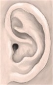
Right auricle, lateral surface 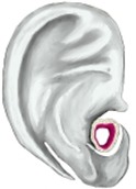
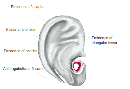
Auricle, cranial (medial) surface
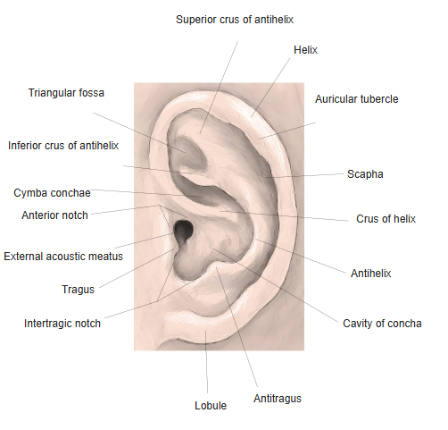
Surface features
| Feature | Description |
|---|---|
| Helix | Curved free rim of the auricle; it begins in the concha as the crus of helix. A small auricular tubercle (Darwin tubercle) may be present on its upper back part |
| Antihelix | Curved ridge in front of the helix; above it divides into the superior and inferior crura, which enclose the triangular fossa |
| Scapha | Groove between the helix and antihelix |
| Concha | Deep hollow in front of the antihelix, divided by the crus of helix into the cymba conchae (above) and the cavity of concha (below), which leads into the meatus |
| Tragus | Small flap in front of the opening of the meatus |
| Antitragus | Small projection opposite the tragus, separated from it by the intertragic notch |
| Lobule | Soft lower part without cartilage |
- On the cranial surface the hollows of the lateral surface appear as raised eminences (of the scapha, concha and triangular fossa).
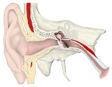
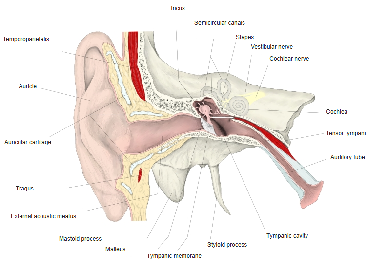
Ear, coronal section
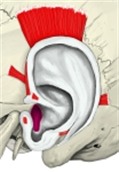
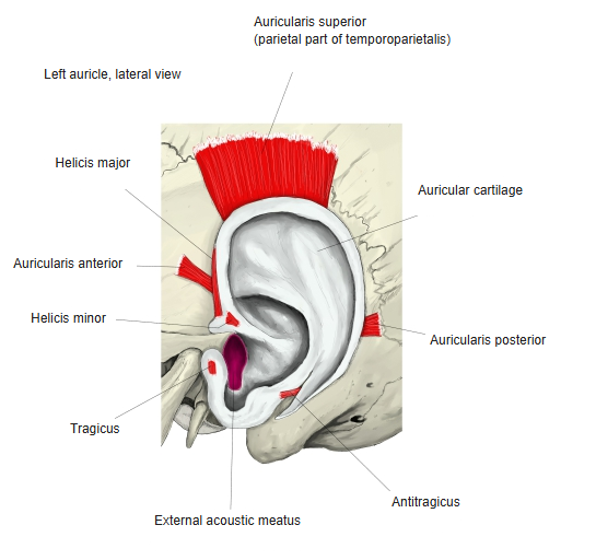
Auricular muscles
- Extrinsic muscles join the auricle to the scalp and skull: auricularis anterior, auricularis superior and auricularis posterior.
- Intrinsic muscles lie on the cartilage itself: helicis major and minor, tragicus, antitragicus, and the transverse and oblique muscles of the auricle.
- All are supplied by the facial nerve; in humans they are rudimentary and move the auricle little or not at all.
Auricular muscles
Blood supply and nerves
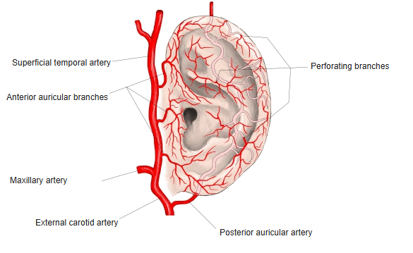
- Arteries: the posterior auricular artery and the anterior auricular branches of the superficial temporal artery; perforating branches pass through the cartilage.
- Veins: follow the arteries to the superficial temporal and posterior auricular veins.
- Lymph: to the preauricular (parotid), mastoid and superficial cervical lymph nodes.
- Sensory nerves: great auricular nerve (most of the cranial surface and lower lateral surface), auriculotemporal nerve (upper front part), lesser occipital nerve (upper cranial surface) and the auricular branch of the vagus nerve (concha).
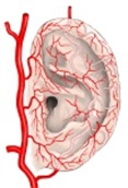
Arteries of the auricle
References and sources
- Further reading: Auricle, Wikipedia.