Latin: meatus acusticus externus
Overview
- The canal of the external ear, running from the concha of the auricle to the tympanic membrane.
- About 2.5 cm long in the adult and slightly S-shaped; to see the tympanic membrane the auricle is pulled upwards and backwards.
- Lateral one-third cartilaginous, medial two-thirds bony (mainly the tympanic part of the temporal bone).
- Conducts sound to the tympanic membrane and protects it. A short definition is also in the glossary: external acoustic meatus.
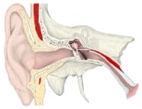
Ear, coronal section
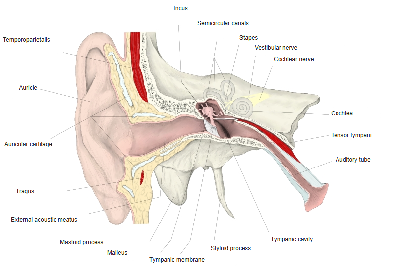
Structure
| Part | Wall | Lining |
|---|---|---|
| Cartilaginous part (lateral third) | Continuation of the auricular cartilage, open above and behind (closed by fibrous tissue) | Thick skin with hairs, sebaceous glands and ceruminous glands (earwax) |
| Bony part (medial two-thirds) | Tympanic part of the temporal bone; the squamous part forms the roof | Thin skin firmly attached to the periosteum, without hairs or glands |
- The narrowest point (isthmus) lies in the bony part, near its junction with the cartilaginous part.
- The tympanic membrane closes the medial end obliquely, so the floor and front wall of the canal are longer than the roof and back wall.
- Earwax and the outward movement of the skin keep the canal clean.
Blood supply and nerves
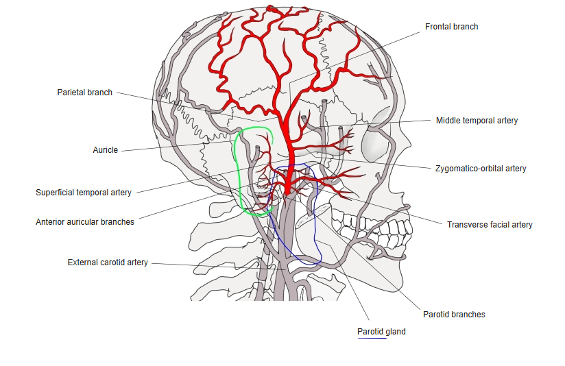
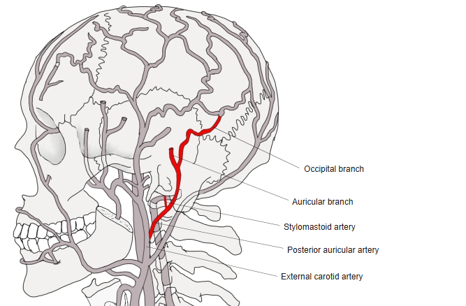
- Arteries: the posterior auricular artery, the deep auricular artery (from the maxillary artery) and the anterior auricular branches of the superficial temporal artery.
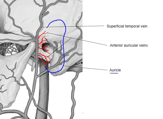
- Veins: to the superficial temporal, posterior auricular and maxillary veins and the pterygoid plexus.
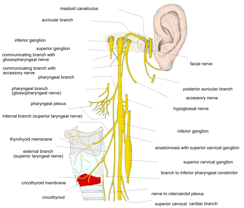
- Nerves: the auriculotemporal nerve supplies the front and roof; the auricular branch of the vagus nerve supplies the back and floor – touching it may cause coughing.
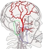
Superficial temporal artery 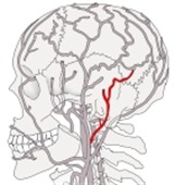
Posterior auricular artery 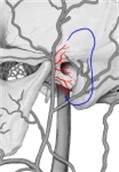
Veins of the auricle 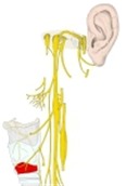
Auricular branch of the vagus nerve
References and sources
- Further reading: External acoustic meatus, Wikipedia.