Latin: tunica vasculosa bulbi
Overview
- The middle coat of the eyeball, rich in blood vessels and pigment; also called the uvea (uveal tract).
- Lies between the fibrous layer outside and the retina inside.
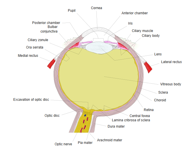
- Three parts from back to front: choroid, ciliary body and iris.
- Nourishes the outer retina, makes the aqueous humor, and controls focusing (ciliary muscle) and the size of the pupil (iris muscles).
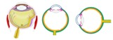
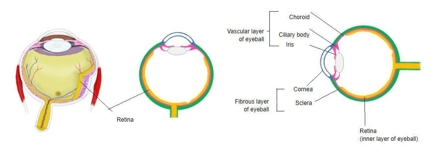
Retina and the layers of the eyeball wall 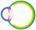
Three layers of the wall (diagram)
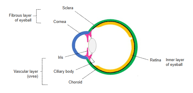
Parts of the vascular layer
| Part | Position | Main features |
|---|---|---|
| Choroid | From the optic nerve to the ora serrata | Thin, dark-brown vascular sheet; supplies the photoreceptors |
| Ciliary body | Ring between the ora serrata and the root of the iris | Ciliary muscle (accommodation) and ciliary processes (aqueous humor); holds the lens by the ciliary zonule |
| Iris | In front of the lens, around the pupil | Pigmented diaphragm with the sphincter and dilator pupillae |
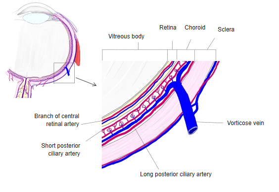
- Arteries: short and long posterior ciliary and anterior ciliary arteries (from the ophthalmic artery); veins: mainly the vorticose veins.
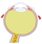
Right eyeball, horizontal section (model) 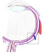
Choroid and its vessels
References and sources
- Further reading: Vascular layer of eyeball, Wikipedia.