Latin: Valva tricuspidalis (valva atrioventricularis dextra)
Overview
- The right atrioventricular valve, between the right atrium and the right ventricle.
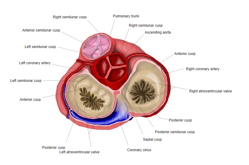
- Has three cusps: anterior, posterior and septal.
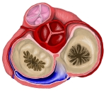
Base of the heart: the four valves seen from above
Structure and function
- The cusps are attached to a fibrous ring of the cardiac skeleton.
- Chordae tendineae link the free edges to the papillary muscles, which tense during ventricular contraction and stop the cusps turning back into the atrium.
- Closes at the start of ventricular systole (part of the first heart sound).
Clinical notes
- Heard best at the lower left sternal border. Tricuspid regurgitation is often secondary to a dilated right ventricle.
References and sources
- Further reading: Tricuspid valve, Wikipedia.