Latin: Valva mitralis (valva atrioventricularis sinistra)
Overview
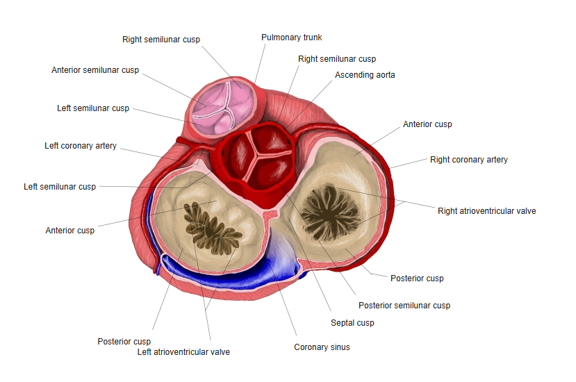
- The left atrioventricular valve (bicuspid valve), between the left atrium and the left ventricle.
- Has two cusps: anterior and posterior.
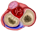
Base of the heart: the four valves seen from above
Structure and function
- The anterior cusp lies between the inflow and the outflow of the left ventricle and is in continuity with the aortic valve.
- Chordae tendineae from two papillary muscles hold the cusps during systole.
- Closes at the start of ventricular systole (part of the first heart sound).
Clinical notes
- Heard best at the apex.
- Mitral stenosis (often after rheumatic fever) enlarges the left atrium; mitral regurgitation and mitral valve prolapse are common valve disorders.
References and sources
- Further reading: Mitral valve, Wikipedia.