Latin: Valva trunci pulmonalis
Overview
- The semilunar valve at the outflow of the right ventricle, at the root of the pulmonary trunk.
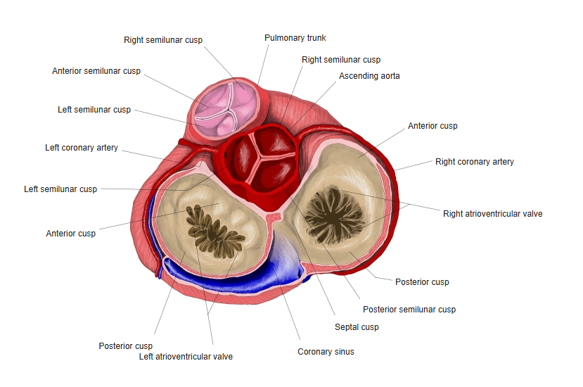
- Has three semilunar cusps: anterior, right and left.
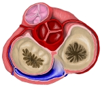
Base of the heart: the four valves seen from above
Structure and function
- Each cusp is a pocket open toward the artery; blood flowing back fills the pockets and closes the valve.
- Closes at the end of ventricular systole (part of the second heart sound).
- Lies the most anterior and superior of the four valves, behind the left 3rd costal cartilage.
Clinical notes
- Heard best in the left 2nd intercostal space. Pulmonary stenosis is usually congenital.
References and sources
- Further reading: Pulmonary valve, Wikipedia.