Latin: Sinus sagittalis superior
Overview
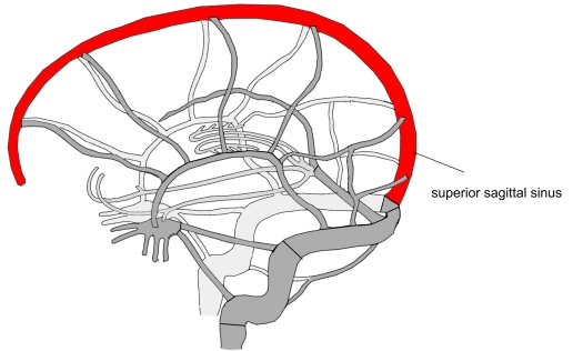
- Unpaired sinus in the attached upper border of the falx cerebri, along the midline of the skull vault.
- Begins near the crista galli (foramen caecum) and runs backward, getting wider as it goes.
- Ends at the internal occipital protuberance, usually turning into the right transverse sinus or joining the confluence of sinuses.
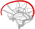
Superior sagittal sinus 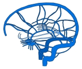
Dural venous sinuses and cerebral veins
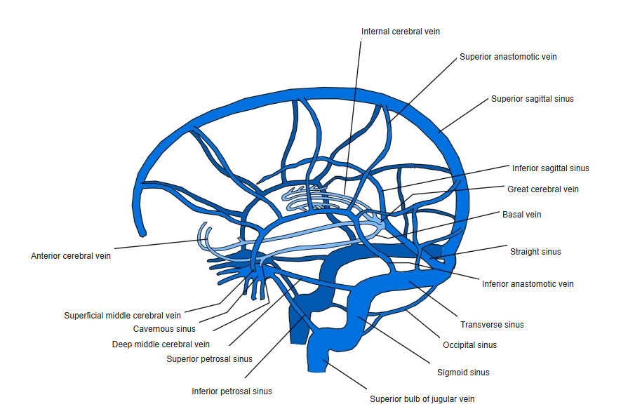
Tributaries
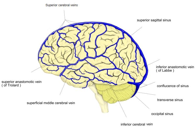
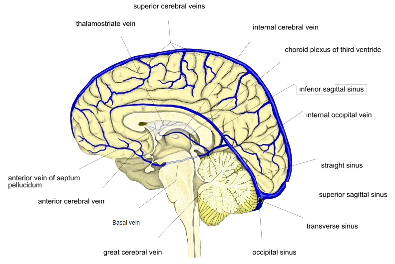
- Superior cerebral veins (they open against the direction of flow).
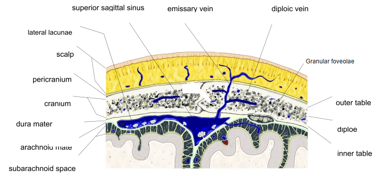
- Diploic and meningeal veins; the parietal emissary veins connect it with veins of the scalp.
- The lateral lacunae open into its sides.
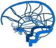
Superficial cerebral veins and dural sinuses 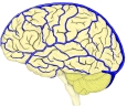
Superficial veins of the lateral surface of the brain 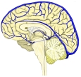
Veins of the medial surface of the brain
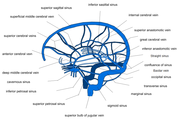
Cerebrospinal fluid
- Arachnoid granulations project into the sinus and its lateral lacunae; cerebrospinal fluid returns to the blood here.
Lateral lacunae and arachnoid granulations
Clinical notes
- The most common site of cerebral venous sinus thrombosis.
References and sources
- Further reading: Superior sagittal sinus, Wikipedia.