Latin: Vena ophthalmica superior
Overview
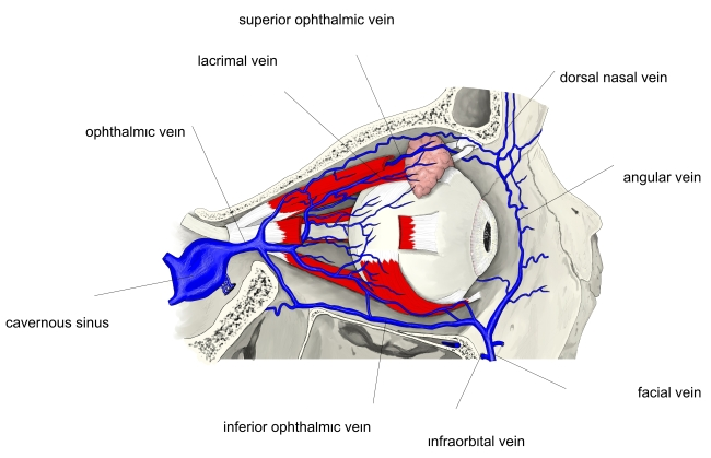
- Main vein of the orbit.
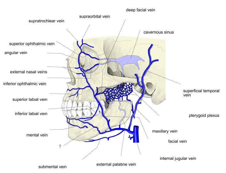
- Begins at the medial angle of the eye by the union of veins that connect with the angular (facial) vein.
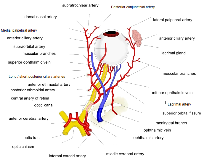
- Runs back with the ophthalmic artery, passes through the superior orbital fissure and ends in the cavernous sinus.
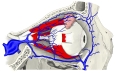
Ophthalmic veins 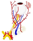
Vessels of the orbit
Tributaries
- Veins matching the branches of the ophthalmic artery: ethmoidal, lacrimal, muscular, vorticose and central retinal veins.
- The inferior ophthalmic vein often joins it.
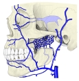
Veins of the face and the cavernous sinus 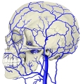
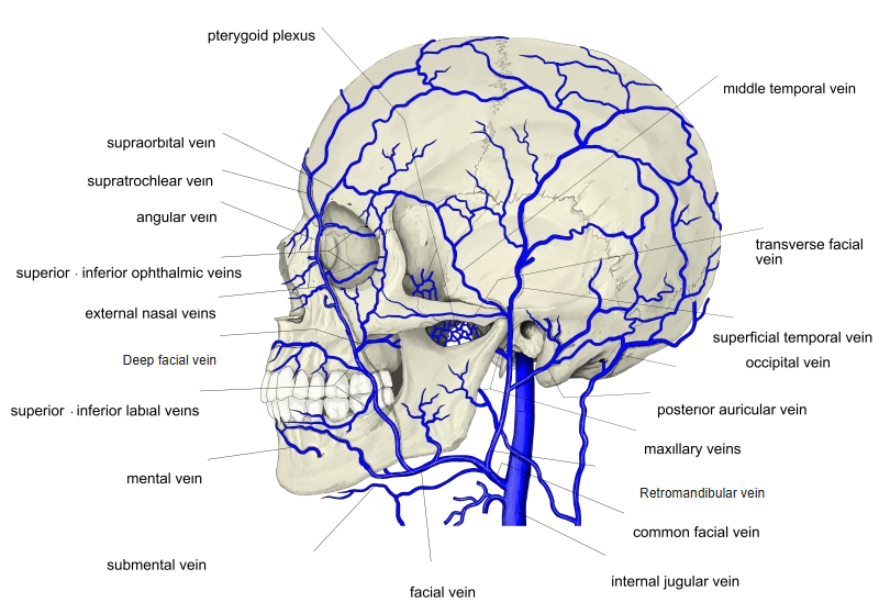
Veins of the head (side view) 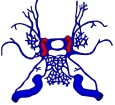
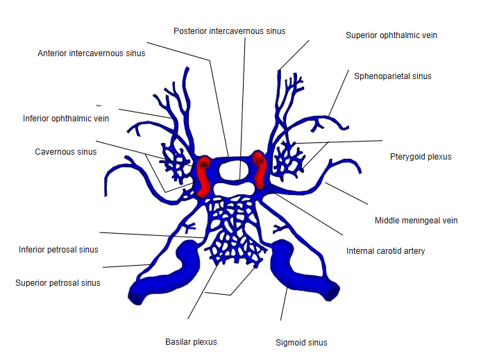
Sinuses around the cavernous sinus 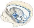
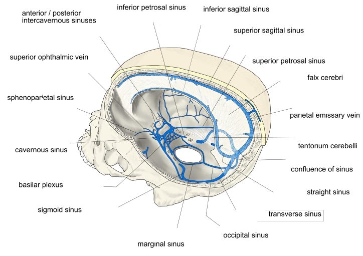
Dural venous sinuses (side view)
Clinical notes
- Its valveless connection between the face and the cavernous sinus is a route for spread of infection.
- It becomes swollen in a carotid–cavernous fistula.
References and sources
- Further reading: Superior ophthalmic vein, Wikipedia.