Superior orbital fissure (Latin: fissura orbitalis superior)
Connects: Orbit ↔ superior orbital fissure ↔ cranial cavity (middle cranial fossa)
It is bounded above by the lesser wing, below by the greater wing and medially by the body of the sphenoid; at its lateral end it is completed by the frontal bone. It is roughly comma-shaped: wide at its medial end and narrowing as it runs upwards and laterally. It lies at the apex of the orbit, just lateral to and below the optic canal, which carries the optic nerve; the foramen rotundum lies just below its medial end.
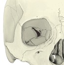 Right orbit (anterior)
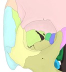 Bones of the right orbit
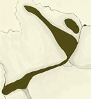 Apex of the right orbit
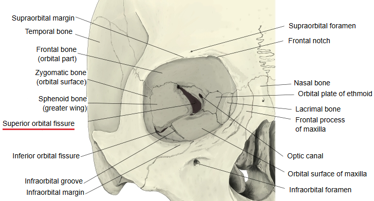
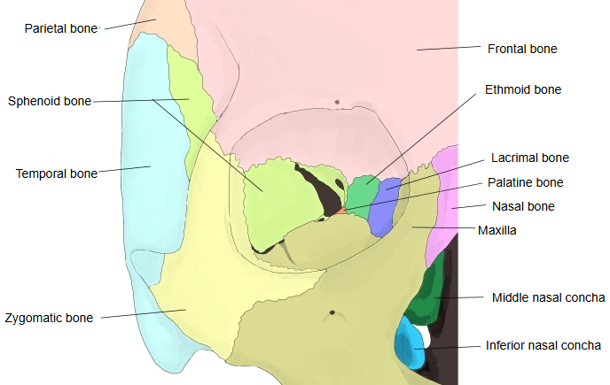
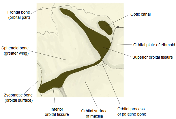
Structures passing through the superior orbital fissure
Several nerves pass through it; the vessel passing through it is the superior ophthalmic vein.
| Nerves | 1 | Abducent nerve (supplies an eye muscle: the lateral rectus) | 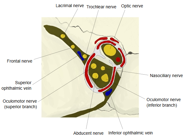 |
| 2 | Trochlear nerve (supplies an eye muscle: the superior oblique) | ||
| 3 | Oculomotor nerve (superior and inferior branches) (supplies the other eye muscles and the levator of the upper eyelid) | ||
| 4 | Lacrimal nerve (branch of the ophthalmic nerve) | ||
| 5 | Frontal nerve (branch of the ophthalmic nerve) | ||
| 6 | Nasociliary nerve (branch of the ophthalmic nerve) | ||
| Vein | Superior ophthalmic vein | ||
Note: some textbooks only say that the ophthalmic nerve passes through, without naming its branches.
Parts of the fissure
The tendinous ring from which the rectus muscles of the eye arise (the common tendinous ring) bridges the fissure and divides it into three parts.
| Part | Structures passing through | |
|---|---|---|
| 1 | Lateral part (above and outside the ring) | Lacrimal nerve, frontal nerve, trochlear nerve (IV), superior ophthalmic vein, and a small anastomotic branch between the lacrimal and middle meningeal arteries |
| 2 | Middle part (inside the ring) | Superior and inferior branches of the oculomotor nerve (III), nasociliary nerve, abducent nerve (VI), and sympathetic fibres |
| 3 | Medial or lower part | The inferior ophthalmic vein (when it does not leave through the inferior orbital fissure) and sympathetic fibres from the plexus around the internal carotid artery |
Sources differ on how the fissure is divided and on which small vessels pass through each part.
Clinical significance
· Fractures, tumours or inflammation at the fissure can damage several cranial nerves at once (superior orbital fissure syndrome).
· Typical signs are paralysis of the eye muscles with double vision, a drooping upper eyelid, a protruding eye and loss of sensation in the forehead.
· If the optic nerve is also affected, with loss of vision, the condition is called orbital apex syndrome.
Reference websites
[Wikipedia]
· Superior orbital fissure, Wikipedia.
[Websites with illustrations]
· Superior orbital fissure: location and contents (Kenhub)
· Left human orbit, numbered openings (Wikimedia Commons)

· Orbit and its openings (Sobotta 1909) (Wikimedia Commons)

Where it is found
Related terms
References and sources
- Further reading: Superior orbital fissure, Wikipedia.
- Further reading: Superior orbital fissure syndrome, Wikipedia.
- Federative International Programme for Anatomical Terminology (FIPAT). Terminologia Anatomica, 2nd ed. (TA2), 2019.