Latin: Sinus cavernosus
Overview
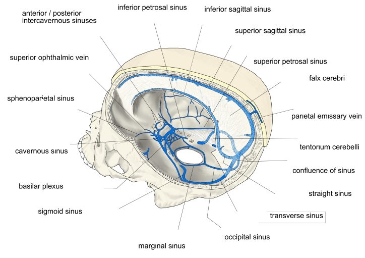
- Paired sinus on each side of the body of the sphenoid and the pituitary fossa.
- Its interior is divided by thin strands into many small spaces (hence "cavernous").
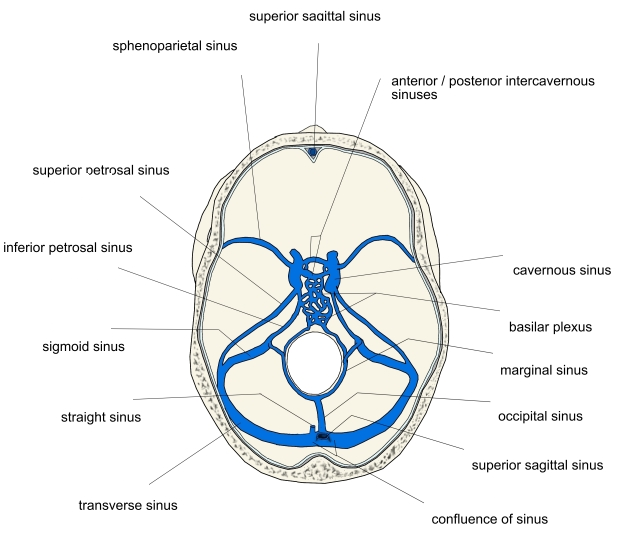
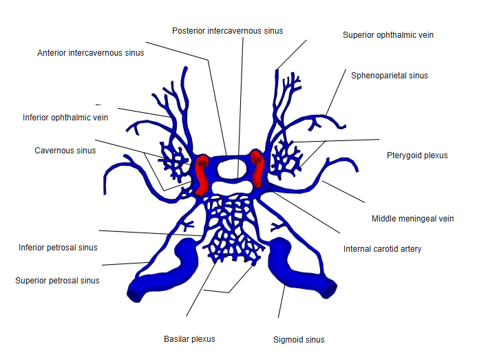
- Joined to the opposite sinus by the anterior and posterior intercavernous sinuses.
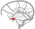
Cavernous sinus 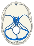
Dural venous sinuses of the cranial base
Tributaries and drainage
- Receives the superior and inferior ophthalmic veins, the superficial middle cerebral vein and the sphenoparietal sinus.
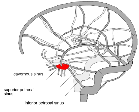
- Drains backward through the superior and inferior petrosal sinuses, and downward to the pterygoid plexus through emissary veins.
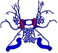
Sinuses around the cavernous sinus 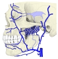
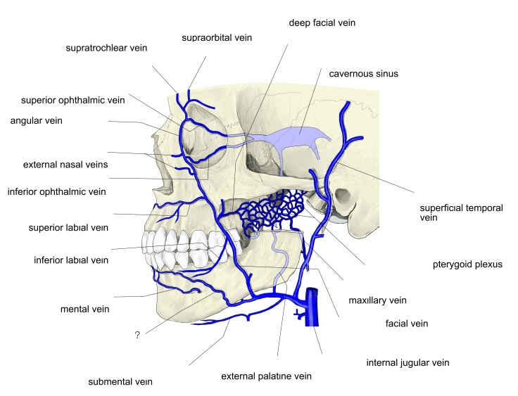
Veins of the face and the cavernous sinus
Structures passing through
- Inside the sinus: the internal carotid artery and the abducent nerve.
- In its lateral wall: the oculomotor and trochlear nerves, and the ophthalmic and maxillary divisions of the trigeminal nerve.
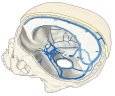
Dural venous sinuses (side view)
Clinical notes
- Infection of the face around the nose and upper lip can reach the sinus through the valveless facial and ophthalmic veins (cavernous sinus thrombosis).
- A tear of the internal carotid artery inside the sinus causes a carotid–cavernous fistula (pulsating bulging eye).
References and sources
- Further reading: Cavernous sinus, Wikipedia.