Interlobular connective tissue (Latin: textus connectivus interlobularis)
Also called the interlobular septa (of lung)
· It is loose connective tissue with collagen and elastic fibres. At the surface it is continuous with the visceral pleura; deeper in, it joins the connective tissue around the bronchi and pulmonary arteries.
· The pulmonary veins and lymphatic vessels at the edge of each lobule run in these septa, while the lobular bronchiole and its artery enter the centre of the lobule.
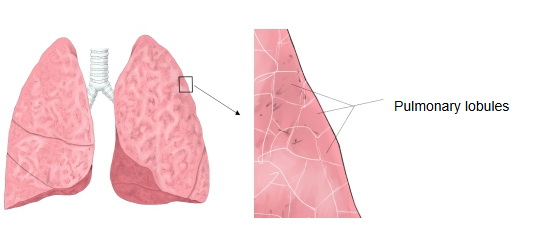
· On the lung surface the septa outline small polygons about 1–2 cm across. In adults, inhaled dust and carbon collect in this tissue, which makes the lobules stand out more clearly.
· The septa are best developed near the surface of the lung and are often incomplete deeper inside.
· When the septa are thickened by fluid, fibrosis or tumour, they become visible on imaging: as short horizontal lines at the lung bases on chest X-rays (Kerley B lines) and as polygonal outlines on high-resolution CT.
Note: the same name is also used for the connective tissue between the lobules of other glands. In the liver it is the connective tissue of the portal canals, around the portal triads.
Where it is found
Related terms
References and sources
- Further reading: Lung, Wikipedia.
- Further reading: Kerley lines, Wikipedia.
- Federative International Programme for Anatomical Terminology (FIPAT). Terminologia Anatomica, 2nd ed. (TA2), 2019.