Pulmonary lobule (Latin: lobulus pulmonis)
Also called the secondary pulmonary lobule or the lobule of lung or the plural lobules
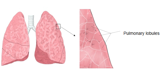
On the surface of each lobe of the lung, small polygonal areas about 1–2 cm across (up to about 2.5 cm) can be seen with the naked eye; these are the pulmonary lobules. Many lobules are bound together by interlobular connective tissue. When much dust and carbon is deposited in this connective tissue, as in adults, the outlines of the lobules become even clearer.
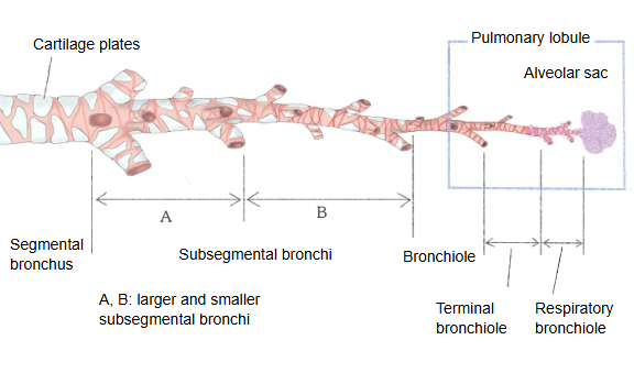
The fine stem that enters a lobule at the end of the segmental bronchial branches is the bronchiole (lobular bronchiole), only about 0.8–1 mm across. Inside the lobule it divides further into 3–6 branches (commonly 3–5) that pass into the next order of airway.
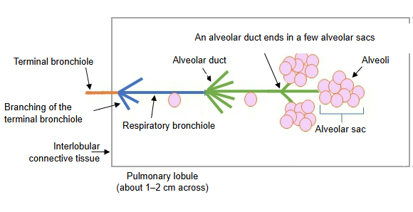
The cone-shaped region, about 1–2 cm across, supplied by one bronchiole is the pulmonary lobule. The bronchiole and a branch of the pulmonary artery enter the centre of the lobule; the pulmonary veins and lymphatics run in the septa at its edge.
Terminal bronchiole: inside the lobule the bronchiole divides into terminal bronchioles. The region supplied by one terminal bronchiole is a pulmonary acinus; within it the airway continues as respiratory bronchioles (with alveoli in their walls), then alveolar ducts, alveolar sacs and alveoli. An alveolar sac is a cluster of a few dozen alveoli.
Sources differ in how they define a lobule: by its connective tissue septa (the usual definition, used here) or by the pattern of bronchiolar branching, and some use "primary lobule" for much smaller units. On high-resolution CT, thickened interlobular septa outline the secondary pulmonary lobules, a useful sign in lung disease.
Reference websites
[Wikipedia]
· Lung, Wikipedia.
· Bronchiole, Wikipedia.
[Websites with illustrations]
· Secondary pulmonary lobule (illustration) (Wikimedia Commons)

· Secondary pulmonary lobule (diagram) (Radiology Key)

· Secondary pulmonary lobule on CT (European Society of Radiology (EPOS))

Where it is found
Related terms
References and sources
- Further reading: Lung, Wikipedia.
- Further reading: Bronchiole, Wikipedia.
- Federative International Programme for Anatomical Terminology (FIPAT). Terminologia Anatomica, 2nd ed. (TA2), 2019.