Alveolar sac (Latin: sacculus alveolaris)

Also called the plural alveolar sacs

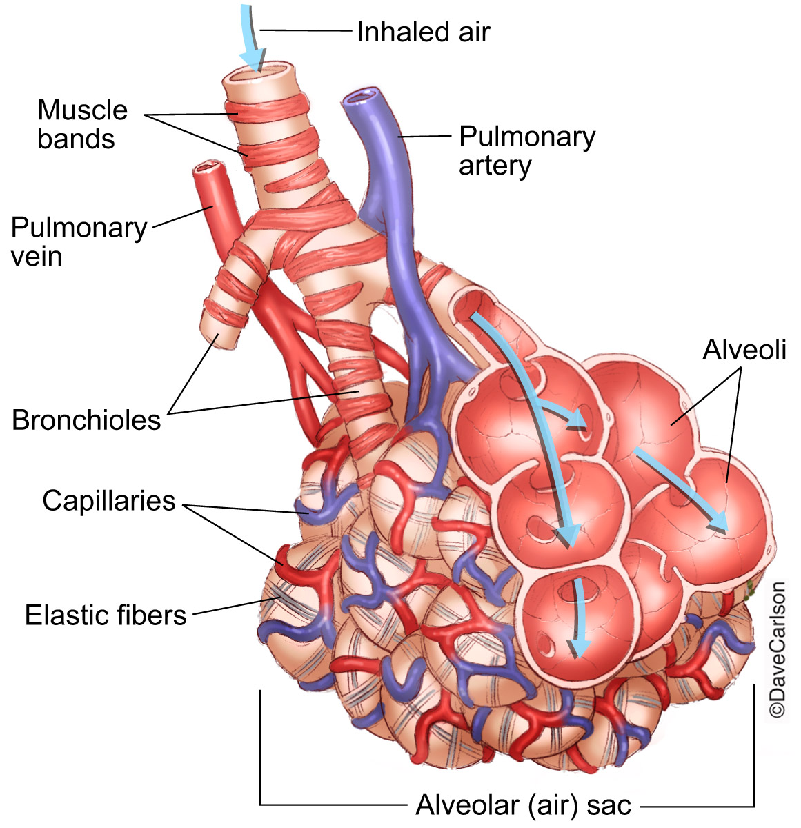
An alveolar sac is the part of the lung at the end of an alveolar duct, where the alveoli crowd together into a blind, bag-like swelling.
Lung: the organ of gas exchangeAlveoli: each about 0.2–0.3 mm across; about 300–500 million in an adult (sources differ)Bag-like swelling: the centre of gas exchange
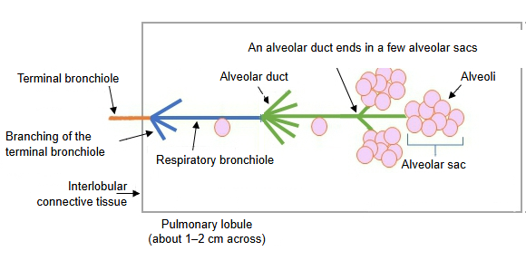
From each alveolar duct, 2–5 alveolar sacs are joined in a grape-like cluster, and the whole lies inside a small compartment called the pulmonary lobule.
2–5: some sources give 5 or 6Pulmonary lobule: about 1–2 cm across, polygonal
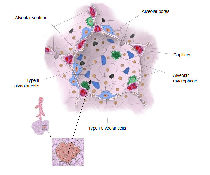
Neighbouring alveoli within an alveolar sac meet at a wall called the alveolar septum and communicate through small holes called alveolar pores.
Alveolar septum: contains capillaries, nerve fibres, elastic fibres and other structuresAlveolar pores: about 5–15 µm across

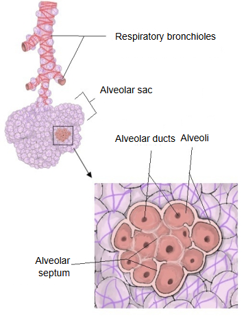
The alveolar sac is the blind, bag-like end of an alveolar duct, divided into several small chambers.

These chambers are the alveoli. Neighbouring alveoli are separated by walls called alveolar septa.

The opening of each alveolus is round. In the free edge of the septum around it, elastic fibres, collagen fibres and a few smooth muscle fibres run in a ring, forming the alveolar ring.

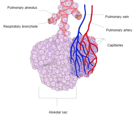
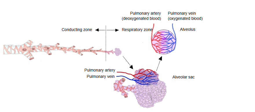
Branches of the pulmonary artery bring deoxygenated blood into the capillary networks around the sacs, and the pulmonary veins carry the oxygenated blood back to the heart (pulmonary circulation). Most gas exchange in the lung takes place here.

From the bronchi to the alveoli
Conducting zone
Main bronchusright or left; enters the lung at the hilum
Lobar bronchiright 3, left 2
Bronchioles (still conducting)
Terminal bronchioleend of the conducting zone
Respiratory zone (gas exchange)
An airway passing from the conducting zone into the respiratory zone, an alveolar sac covered by branches of the pulmonary artery and vein, and an enlarged alveolus with its capillary network.
From airway to alveolus (click to enlarge)
A schematic pulmonary lobule: a terminal bronchiole, a respiratory bronchiole, alveolar ducts and alveolar sacs made of alveoli.
Pulmonary lobule (schematic)
  • An alveolus seen from inside, with alveolar septa, alveolar pores, capillaries, an alveolar macrophage and type I and type II alveolar cells.Alveolar wall
  • Respiratory bronchioles leading to an alveolar sac, with an enlarged view of a cluster of alveoli separated by alveolar septa.Respiratory bronchioles and alveolar sac
  • An alveolar sac at the end of a respiratory bronchiole, partly covered by a capillary network between branches of the pulmonary artery and pulmonary vein.Alveolar sac and blood vessels

Reference websites

[Wikipedia]

· Pulmonary alveolus, Wikipedia.

· Alveolar duct, Wikipedia.

[Websites with illustrations]

· Alveolar sac (IMAIOS e-Anatomy)

· Diagram of alveoli (GeeksforGeeks)

· Bronchioles and alveoli (illustration) (Carlson Stock Art)
Where it is found

References and sources

  1. Further reading: Pulmonary alveolus, Wikipedia.
  2. Further reading: Alveolar duct, Wikipedia.
  3. Federative International Programme for Anatomical Terminology (FIPAT). Terminologia Anatomica, 2nd ed. (TA2), 2019.