Left lobe of liver (Latin: lobus hepatis sinister)
Also called the left lobe or the left liver (functional)
into right and left
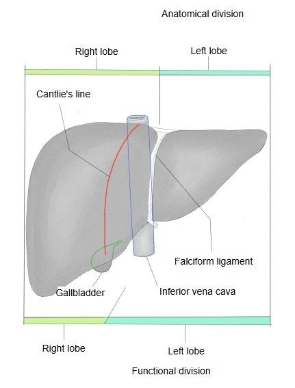
- Anatomical division : by the surface of the liver. The boundary is the attachment of the falciform ligament. The right lobe makes up about three-quarters to four-fifths of the liver, the left lobe the rest.
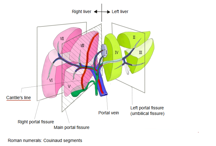
- Functional division : by the branching of the vessels and bile ducts. The boundary is Cantlie's line (the main portal fissure). The right liver makes up about 60-65% of the liver. The caudate lobe is treated as independent: it belongs to neither the right nor the left liver.
· The left lobe is thinner and flatter than the right lobe. It extends to the left across the epigastrium into the left hypochondrium, over the stomach. Its pointed left end tapers into the fibrous appendix of the liver, a band of fibrous tissue at the end of the left triangular ligament.
· Anatomically it lies to the left of the falciform ligament on the diaphragmatic surface, and to the left of the fissure for the ligamentum teres and the fissure for the ligamentum venosum on the visceral surface. Its visceral surface bears the gastric impression and, near the top, the small esophageal impression; a rounded bulge, the omental tuberosity, faces the lesser omentum. It is attached to the diaphragm by the left part of the coronary ligament, which ends as the left triangular ligament.
· The two left sides do not coincide: the functional left liver lies to the left of Cantlie's line (from the gallbladder fossa to the inferior vena cava), so it also includes the quadrate lobe and the region above it. It is supplied by the left branches of the portal vein and hepatic artery and drained by the left hepatic duct; the left hepatic vein drains most of it into the inferior vena cava.
· The umbilical fissure, marked by the falciform ligament and ligamentum teres, divides the left liver into a medial section (segment IV, with IVa above and IVb, the quadrate lobe, below) and a lateral section (segments II and III) of the Couinaud hepatic segments. The lateral section is the part removed in a left lateral sectionectomy, for example in living donor liver transplantation to a child.
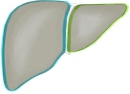 Anatomical division
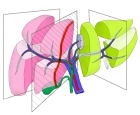 Functional division
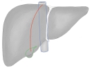 Cantlie's line
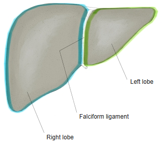
Where it is found
Related terms
- Right lobe of liver
- Quadrate lobe
- Caudate lobe
- Hepatic segments
- Fissure for ligamentum teres
- Fissure for ligamentum venosum
- Liver
References and sources
- Further reading: Lobes of liver, Wikipedia.
- Further reading: Liver segment, Wikipedia.
- Federative International Programme for Anatomical Terminology (FIPAT). Terminologia Anatomica, 2nd ed. (TA2), 2019.