Caudate lobe (Latin: lobus caudatus)
Also called Spigelian lobe or the segment I (Couinaud)
Anatomical division
right lobe
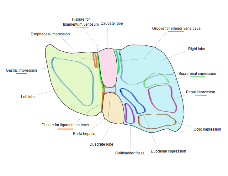
- Right lobe (in the narrow sense) : the part of the right lobe apart from the quadrate and caudate lobes.
- Quadrate lobe : below and in front; a four-sided area bounded by the fissure for the ligamentum teres, the gallbladder fossa and the porta hepatis.
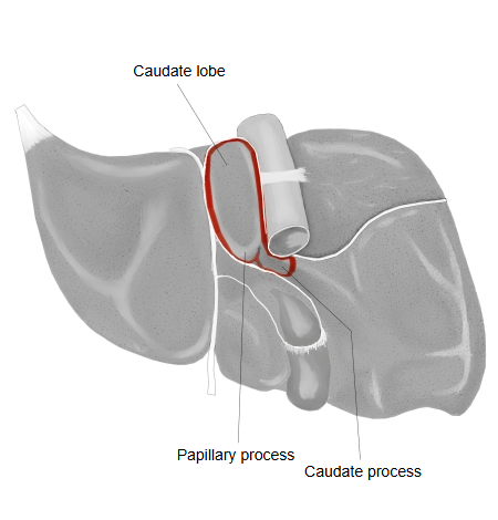
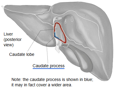
- Caudate lobe : behind; bounded by the groove for the inferior vena cava, the fissure for the ligamentum venosum and the porta hepatis.
Hepatic segments
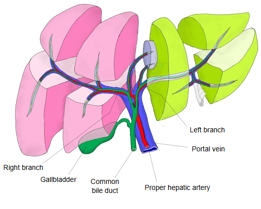
Viewed functionally, the caudate lobe belongs to neither the right lobe nor the left lobe and is regarded as independent. It receives branches from both the right and left branches of the portal vein and hepatic artery, and its bile drains to both hepatic ducts. It drains by several small short hepatic veins straight into the inferior vena cava, not through the main hepatic veins; for this reason it is often spared and enlarged in Budd–Chiari syndrome and in cirrhosis.
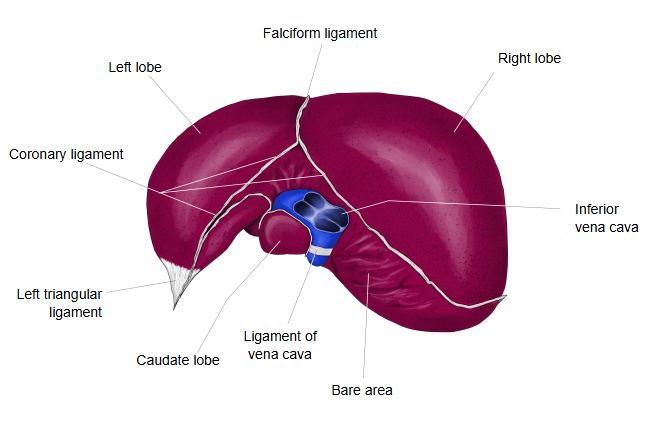
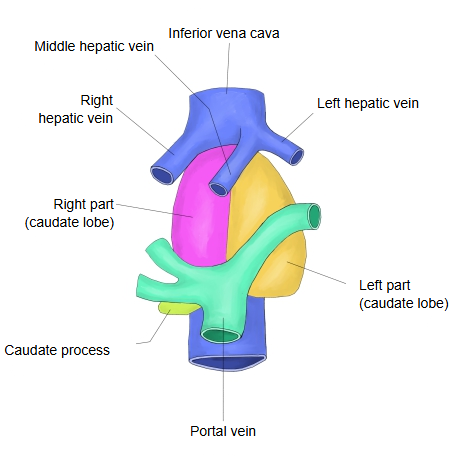
The Healey and Schroy classification divides the caudate lobe into three parts: a right part, a left part and the caudate process. Surgeons often speak of a left part (the Spigelian lobe), a paracaval portion in front of the inferior vena cava and the caudate process; sources differ on the names and boundaries of these parts. Behind the inferior vena cava, the caudate lobe and right lobe are often linked by a fibrous band or a bridge of liver tissue, the ligament of the inferior vena cava (hepatocaval ligament).
(Healey and Schroy)
- Right lobe
- Anterior segment : superior area (Couinaud VIII) and inferior area (Couinaud V).
- Posterior segment : superior area (Couinaud VII) and inferior area (Couinaud VI).
- Caudate lobe : left part, right part and caudate process (Couinaud I).
- Left lobe
- Medial segment : superior area (Couinaud IVa) and inferior area (Couinaud IVb, the quadrate lobe).
- Lateral segment : superior area (Couinaud II) and inferior area (Couinaud III).
· Because it lies behind the porta hepatis, the caudate lobe has also been called the dorsal sector of the liver. In the Couinaud classification it is the independent segment I. It faces the superior recess of the omental bursa, behind the lesser omentum.
Processes
caudate lobe
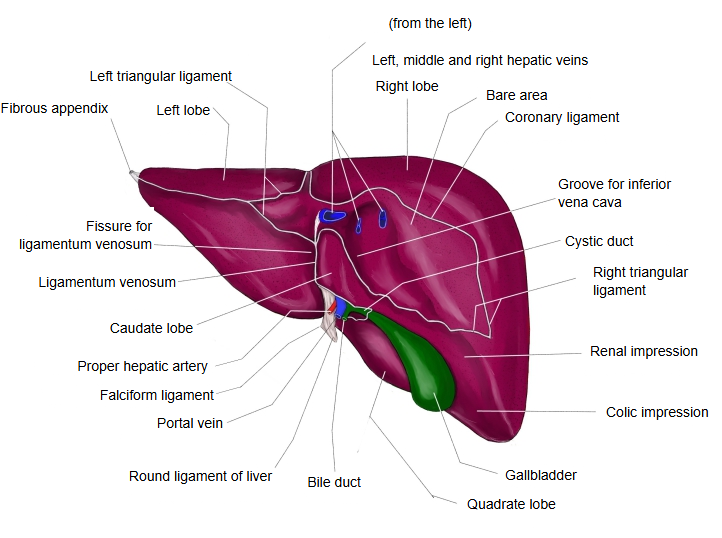
- Papillary process : a rounded knob at the lower left corner that projects down and forward towards the lesser omentum. An enlarged papillary process can be mistaken for an enlarged lymph node or a mass on imaging.
- Caudate process : a narrow ridge of liver tissue behind and above the porta hepatis, below the groove for the inferior vena cava, that joins the caudate lobe to the right lobe. It forms the upper boundary of the omental (epiploic) foramen.
 Caudate lobe (visceral surface)
 Caudate lobe (posterior view)
Size
1. Length: it varies between people, but its vertical length is generally about 3-7 cm (rough estimate).
2. Share of the liver: it usually makes up only about 1-3% of the whole liver (rough estimate).
- Caudate lobe (visceral surface)
- Caudate lobe (posterior view)
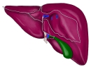 Liver (posterior and visceral surfaces)
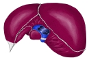 Liver (posterosuperior view)
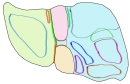 Features of the visceral surface
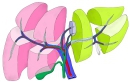 Hepatic segments and portal triad
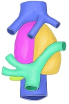 Parts of the caudate lobe
Reference websites
[Wikipedia]
· Caudate lobe of liver, Wikipedia.
· Lobes of liver, Wikipedia.
[Websites with illustrations]
· Liver segments (Couinaud classification) (Radiopaedia)
· Gray's Anatomy: posterior and inferior surfaces of the liver (Wikimedia Commons)

· The liver (TeachMeAnatomy)
Where it is found
Related terms
- Caudate process
- Papillary process
- Porta hepatis
- Fissure for ligamentum venosum
- Groove for vena cava
- Hepatic segments
- Liver
References and sources
- Further reading: Caudate lobe of liver, Wikipedia.
- Further reading: Lobes of liver, Wikipedia.
- Federative International Programme for Anatomical Terminology (FIPAT). Terminologia Anatomica, 2nd ed. (TA2), 2019.