Bronchial glands (Latin: glandulae bronchiales)
Also called the singular bronchial gland
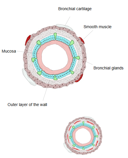
Note: the figure is a simplified drawing; the glands are shown as small flask-shaped bodies, while in the real wall the main parts of the glands lie deeper, in the submucosa.
They are mixed (seromucous) glands in the submucosa. They are fewer than the tracheal glands, become sparse in the smaller bronchi and are absent in the bronchioles, roughly where the cartilage ends.
· The gland bodies lie in the submucosa, mainly outside the smooth muscle and between the cartilage plates; their ducts pass inward to open on the surface of the epithelium.
· They contain both mucous cells and serous cells. The mucus traps dust and microbes, the cilia sweep it up toward the throat (mucociliary clearance), and the serous fluid carries antimicrobial substances such as lysozyme and lactoferrin.
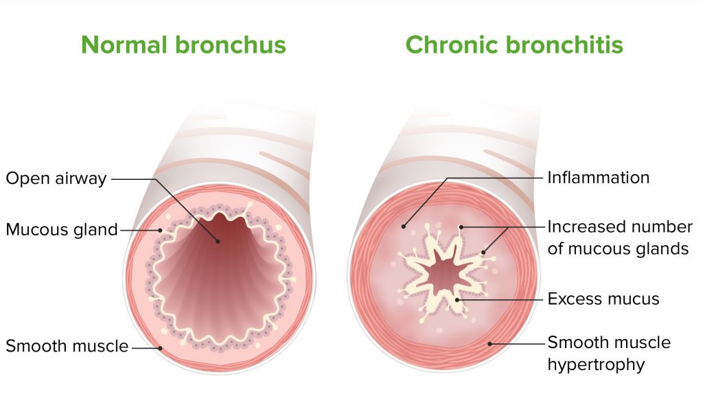
· Parasympathetic (vagal) fibres, through the bronchial branches of the vagus nerve, increase their secretion. Enlarged glands with too much mucus are a feature of chronic bronchitis.
Reference websites
[Wikipedia]
· Bronchus, Wikipedia.
· Mucociliary clearance, Wikipedia.
[Websites with illustrations]
· Bronchiectasis (illustration) (Healthline)

· Bronchiectasis (MSD Manuals)

· Chronic bronchitis pathophysiology (Lifeline Laboratory)
Where it is found
Related terms
References and sources
- Further reading: Bronchus, Wikipedia.
- Further reading: Mucociliary clearance, Wikipedia.
- Federative International Programme for Anatomical Terminology (FIPAT). Terminologia Anatomica, 2nd ed. (TA2), 2019.