Cilia (Latin: cilia)
Also called the cilium (singular)
- Overview
- Microvilli, villi and cilia compared
- Reference websites
- (Wikipedia, websites with illustrations)
- Related terms
Overview
Motile cilia
The places where motile cilia are found and their work are listed below.
| Organ | Work of the cilia | |
|---|---|---|
| 1 | Airway epithelium (trachea, bronchi; a pseudostratified ciliated epithelium) | Carry mucus and foreign matter toward the mouth (= mucociliary clearance) |
| 2 | Epithelium of the uterine tube | Move the egg toward the uterus |
| 3 | Lining of the brain ventricles (ependymal cells) | Help the circulation of the cerebrospinal fluid (CSF) |
| 4 | Efferent ductules of the testis | Help to move the sperm (the duct of the epididymis itself carries stereocilia, which are long microvilli, not cilia) |
· Motile and non-motile cilia differ in their internal structure, but from outside they can hardly be told apart.
| Kind | Microtubules | Motility | Main places | |
|---|---|---|---|---|
| 1 | Motile cilia | 9+2 pattern | yes | Airway, uterine tube and others |
| 2 | Non-motile (primary) cilia | 9+0 pattern | no | Usually one on each of many cells; involved in sensing |
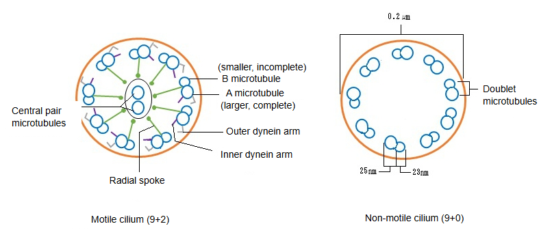
Motile cilia (9+2 pattern): nine outer microtubule doublets surround a central pair; each doublet has a complete A microtubule and an incomplete B microtubule. Dynein arms on the doublets make the cilium beat, and radial spokes link the doublets to the central pair.
Primary cilia (9+0 pattern): nine outer doublets with no central pair and no dynein arms. They act as antennae that sense chemical and mechanical signals; modified cilia form parts of the light receptors of the retina and of the olfactory receptor cells (see olfactory vesicle).
Examples of cells with no cilia at all
1. 1. Red blood cells (mature mammalian red blood cells)
· They have lost their nucleus and organelles, and they have no cilia.
· Being specialized for carrying oxygen, they need no projections for sensing or movement.
2. 2. Platelets
· Cell fragments without a nucleus, specialized for stopping bleeding in the blood vessels.
· They have no cilia.
3. 3. Mature skeletal muscle and cardiac muscle cells
· Specialized for contraction (skeletal muscle fibres are multinucleated), they have almost no primary or motile cilia.
· A primary cilium may be present for a time early in development, but it is lost as the cells mature.
4. 4. Mature keratinocytes (outermost layer of the epidermis)
· The cells of the stratum corneum of the epidermis (keratinocytes) have lost their nucleus and organelles, and they have no cilia.
· They are specialized as a barrier.
In short, cells without a nucleus or organelles, and cells specialized for one function (such as contraction or carrying oxygen), tend to have no cilia.
Microvilli, villi and cilia compared
| Structure | Size and shape | Function | Typical places | |
|---|---|---|---|---|
| 1 | Microvilli | Tiny projections of a cell (seen with the electron microscope); diameter 0.08–0.1 µm, length about 1–2 µm | Absorption, secretion | Small intestine, kidney (tubules) |
| 2 | Villi | Large finger-like projections of the mucosa (seen with the naked eye or light microscope); diameter 0.1–0.3 mm, length 0.5–1.5 mm | Absorption | Small intestine |
| 3 | Cilia | Movable projections of a cell; diameter about 0.2 µm, length about 5–10 µm | Moving mucus and other material | Airway epithelium, uterine tube epithelium |
Reference websites
[Wikipedia]
· Cilium, Wikipedia.
· Axoneme, Wikipedia.
[Websites with illustrations]
· Cilia and flagella (Molecular Expressions (Florida State University))
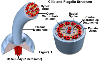
· Structure of cilia (figure) (Elsevier)

· Structure of the cilium (figure) (Springer Nature)

Related terms
- Mucus
- Olfactory vesicle
- Microvilli
- Villi
- Pseudostratified epithelium
- Trachea
- Uterine tube
- Cerebral ventricles