Trachea (Latin: trachea)
- Overview
- Structure (mucosa)
- Parts
- Relations to surrounding structures
- Vessels and nerves
- Related terms
- References
Overview
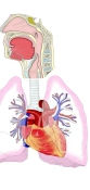
system
- Airway
- Nasal cavity : from the nostrils to the choanae; divided into right and left halves by the nasal septum and lined with mucosa.
- Pharynx : made of three parts (upper, middle, lower) and lined with mucosa; also part of the digestive system.
- Larynx : funnel-shaped tube at the level of the 4th–7th cervical vertebrae; its framework is cartilage.
- Trachea : tube about 10 cm long and 20–25 mm wide; divides at the level of the 4th–5th thoracic vertebrae.
- Bronchi : branch from the trachea to the right and left, and divide into many bronchial branches inside the lungs.
- Lungs : right lung (upper, middle, lower lobes), left lung (upper, lower lobes); gas exchange (external respiration) takes place in the alveoli.
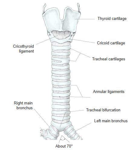
· In the adult the bifurcation lies at about the level of the 2nd costal cartilage (the sternal angle); in small children it is a little lower, at about the level of the 3rd costal cartilage.
· At the bifurcation a ridge projects into the lumen of the trachea; it is called the carina of the trachea.
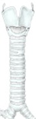 Thyroid cartilage to bifurcation
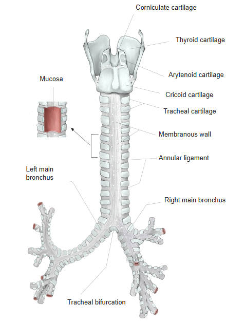
(anterior view)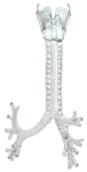 Thyroid cartilage to bronchi
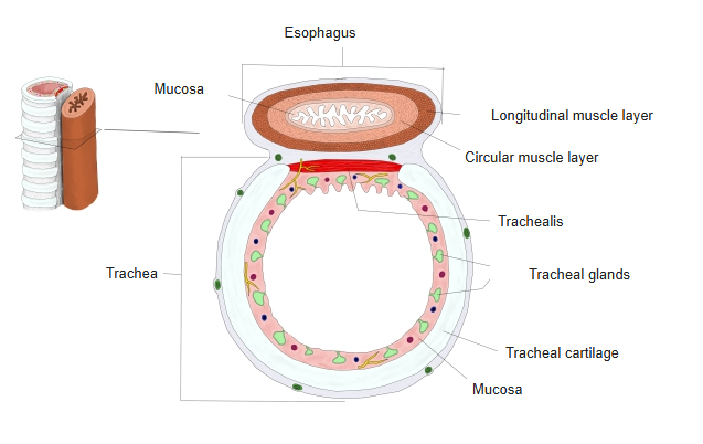
(posterior view)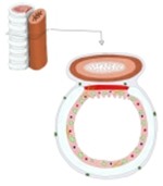 Trachea and esophagus
(cross-section)
Structure
the trachea
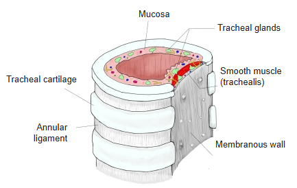
- Tracheal cartilages : 15–20 C-shaped hyaline cartilages, each about 3–4 mm high.
- Annular ligaments : the connective tissue membrane (fibroelastic membrane) around the cartilages, continuing between them.
- Mucosa : well developed and lines the lumen; it forms no folds. Contains tracheal glands, vessels and nerves.
- Membranous wall : the back wall facing the esophagus; made mainly of two layers of smooth muscle (the trachealis), with large tracheal glands.
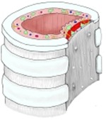 Structure of the trachea
[Mucosa]
The mucosa is well developed but forms no folds.
| 1 | Epithelium | Pseudostratified ciliated columnar epithelium with goblet cells (as in the larynx); the cilia beat upwards. |
| 2 | Basement membrane | Distinct. The lamina propria is rich in elastic fibres, and the submucosa contains many tracheal glands (mixed glands). |
The trachea is made of 15–20 rings of cartilage stacked one above another, the annular ligaments between them, and the mucosa that lines their inner surface. The cartilage rings are called tracheal cartilages and are made of hyaline cartilage. They are not complete rings: they lack their back part (about two-fifths to one-third of the circumference) and so are C-shaped. The back wall, called the membranous wall, is formed by smooth muscle that joins the two ends of each C. The membranous wall is thought to allow the esophagus to widen when food passes through it. The cartilages as a whole are wrapped in a connective tissue membrane rich in elastic fibres (fibroelastic membrane), which also fills the gaps between neighbouring cartilages as the annular ligaments.
The mucosa is well developed and forms no folds. Its epithelium is pseudostratified ciliated columnar epithelium with goblet cells, as in the larynx, and the cilia beat upwards. The basement membrane is distinct, the lamina propria is rich in elastic fibres, and the submucosa contains many tracheal glands (mixed glands), especially large ones in and around the smooth muscle layer of the membranous wall. The muscle layer of the membranous wall has an inner circular and an outer longitudinal layer, together called the trachealis.
Parts
The trachea is broadly divided into a cervical part and a thoracic part.
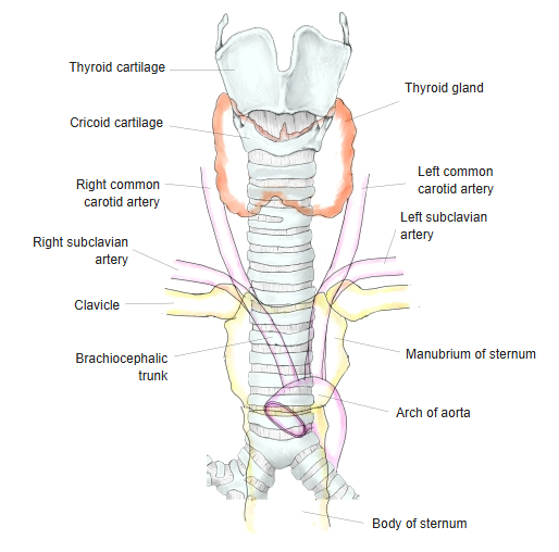
| 1 | Cervical part | Follows the cervical curve of the vertebral column and is slightly convex backwards. Above, it lies superficially in the midline of the neck; lower down it lies deeper and deeper. On its way it is in contact with: 1. Thyroid gland: covers the front and sides of the 2nd–5th tracheal cartilages. 2. Thyroid venous plexus (inferior thyroid veins): lies in front of the lower part of the cervical trachea, sometimes with the thyroid ima artery. 3. Recurrent laryngeal nerve: in the groove between the trachea and the esophagus (tracheoesophageal groove). 4. Vagus nerve: lateral to the cervical part. |
| 2 | Thoracic part | Runs parallel to the vertebral column behind the manubrium of the sternum and the left brachiocephalic vein, at the border between the middle and posterior mediastinum. The brachiocephalic trunk runs on its right and the left common carotid artery on its left. In young people it is separated from the sternum by the thymus, which is well developed at that age. Its back is in contact with the front wall of the esophagus. |
Relations to surrounding structures
The back of the trachea (the membranous wall) is in contact with the esophagus. In the neck the esophagus extends slightly beyond the left edge of the trachea, and lower down it crosses the left main bronchus. The isthmus of the thyroid gland lies in front of the 2nd–5th tracheal cartilages, and the left and right lobes of the gland embrace the upper part of the trachea from both sides.
 Trachea and its surroundings
Vessels and nerves
The vessels and nerves of the trachea are as follows.
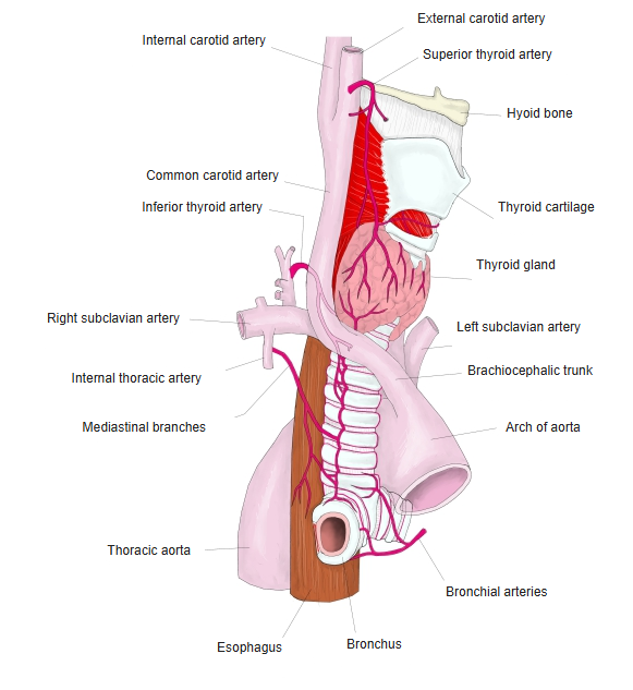
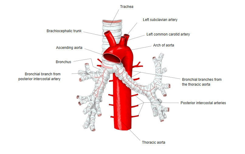
| Arteries | Upper part: tracheal branches of the inferior thyroid artery. Back: bronchial arteries (visceral branches of the thoracic aorta). Front: mediastinal branches of the internal thoracic artery. The branches join with those above and below, forming a network in the tracheal wall. |
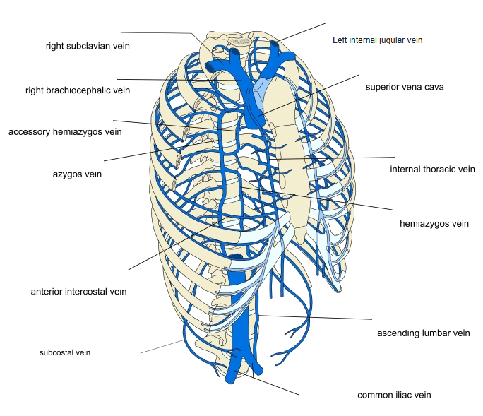
| Veins | Form a network around the trachea that drains into the brachiocephalic veins and the azygos vein. Upper part: inferior thyroid veins. Back: bronchial veins. Front: small veins to the internal thoracic veins. |
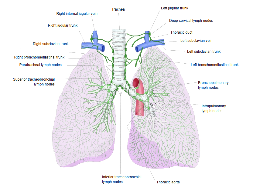
| Lymphatics | Numerous in the mucosa, with lymphocytes in the epithelium. They drain to the pretracheal and paratracheal lymph nodes beside the trachea and to the tracheobronchial nodes around the bifurcation. |
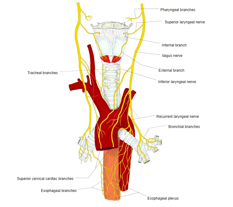
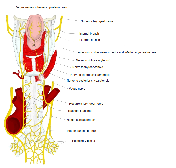
| Nerves | Direct branches of the vagus nerve, the recurrent laryngeal nerve and the inferior laryngeal nerve, together with sympathetic branches. |
 Arteries 1
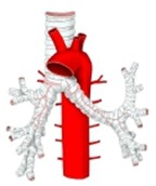 Arteries 2
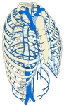 Veins
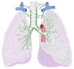 Lymph nodes and lymph trunks
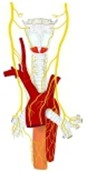 Nerves 1
 Nerves 2
Related terms
Main anatomical terms related to the trachea, in alphabetical order.
References and sources
The trachea is supplied above by the inferior thyroid arteries and drained by the inferior thyroid veins, while its lower part receives blood from the bronchial arteries; these branches meet in the wall of the trachea. Its lymph drains to the pretracheal and paratracheal lymph nodes.
- Further reading: Trachea, Wikipedia.