Bronchi (Latin: bronchi)
Overview
system
- Airway
- Nasal cavity : from the nostrils to the choanae; divided into right and left halves by the nasal septum and lined with mucosa.
- Pharynx : made of three parts (upper, middle, lower) and lined with mucosa; also part of the digestive system.
- Larynx : funnel-shaped tube at the level of the 4th–7th cervical vertebrae; its framework is cartilage.
- Trachea : tube about 10 cm long and 20–25 mm wide; divides at the level of the 4th–5th thoracic vertebrae.
- Bronchi : branch from the trachea to the right and left, and divide into many bronchial branches inside the lungs.
- Lungs : right lung (upper, middle, lower lobes), left lung (upper, lower lobes); gas exchange (external respiration) takes place in the alveoli.
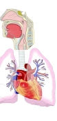
[Right and left main bronchi]
The right and left main bronchi differ in width, length and course.
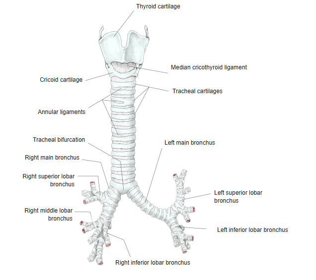
| Left main bronchus | · Makes an angle of about 46° with the midline. · Narrower than the right and about twice as long (about 4–6 cm). · Runs below the aortic arch and in front of the esophagus and the thoracic aorta to the left hilum. · Divides into 2 branches (superior and inferior) and enters the lung at about the level of the 6th thoracic vertebra. · Bronchial cartilages: 7–11. |
| Right main bronchus | · Runs more nearly vertical than the left. · Makes an angle of about 24° with the midline. · Wider and shorter than the left (about 3 cm). · Runs downwards and outwards behind the superior vena cava to the right hilum. · Divides into 3 branches (superior, middle and inferior) and enters the lung at about the level of the 5th thoracic vertebra. · Bronchial cartilages: 3–4. |
Because the two main bronchi leave the bifurcation on different courses, inhaled foreign bodies most often enter the right bronchus. In small children the angle between the two bronchi is about 110°, and the right and left angles do not differ.
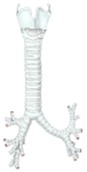 Thyroid cartilage to bronchi
(anterior view)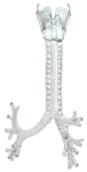 Thyroid cartilage to bronchi
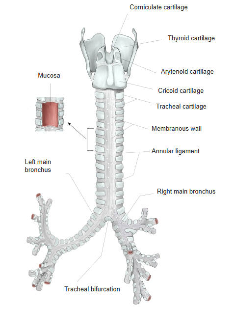
(posterior view) Segmental bronchus
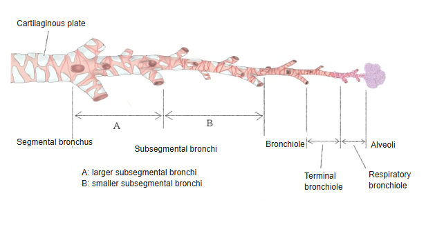
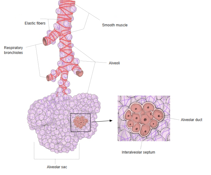
to alveolar sacs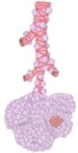 End of the bronchial tree
Structure
the bronchi
- Bronchial cartilages : hyaline cartilage as in the trachea, but they gradually become irregular plates and disappear towards the ends of the tree.
- Annular ligaments : continue from the trachea; where the cartilages become irregular, these ligaments can no longer be told apart.
- Mucosa : well developed and lines the lumen; it forms no folds. The bronchial glands are fewer than the tracheal glands.
- Membranous wall : continues from the trachea; as the cartilages lose their C shape it is no longer a separate back wall.
· The bronchial wall is normally 10–20% as thick as the whole diameter of the bronchus.
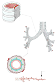 Structure
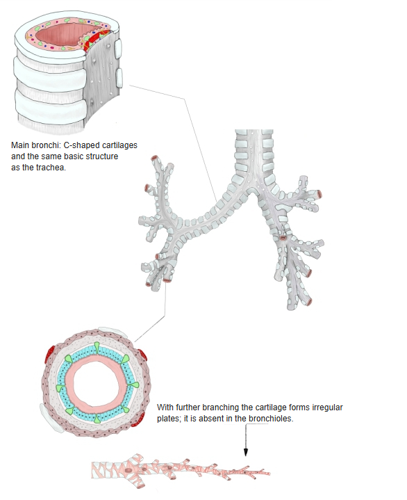
Parts
Inside the lungs the main bronchi divide again and again. The levels of branching are as follows.
| 1 | Lobar bronchi | Branch from the main bronchi at the hilum (2 on the left, 3 on the right). Inside the lung their structure is almost the same as that of the main bronchi, with some differences: · They have no membranous wall. · The C-shaped cartilages gradually shrink into irregular plates (found down to branches about 1 mm wide). · The cells of the pseudostratified ciliated columnar epithelium gradually become lower. |
| 2 | Segmental bronchi | · Each lobar bronchus divides into 2–5 segmental bronchi. · The region supplied by the branches of one segmental bronchus is a bronchopulmonary segment; the segments have fixed numbers. · Irregular cartilage plates are present. |
| 3 | Bronchioles | · The segmental bronchi divide into many segmental bronchial branches. · At the end of these branches, the thin stem that enters a pulmonary lobule is a bronchiole (0.8–1 mm wide). · Within the lobule it divides into 3–6 smaller branches. The cone-shaped region, 1–2 cm across, supplied by one bronchiole is the pulmonary lobule. In the wider bronchioles the epithelium is pseudostratified ciliated epithelium with goblet cells, the lamina propria contains bronchial glands (mixed glands), smooth muscle runs beneath it, and below the muscle is a layer of loose connective tissue with cartilage plates. When a bronchiole becomes narrower than 1 mm, cartilage disappears and the smooth muscle becomes stronger and runs circularly. Excessive contraction of this muscle narrows the airways, as in bronchial asthma. |
| 4 | Terminal bronchioles | · Within the pulmonary lobule the bronchioles divide into terminal bronchioles. · Their epithelium is simple columnar, made of ciliated cells and secretory cells, with a thin layer of scattered smooth muscle beneath it. |
| 5 | Respiratory bronchioles | · Alveoli open from their walls here and there. · Each respiratory bronchiole gives rise to 2–9 alveolar ducts, which end in blind alveolar sacs surrounded by alveoli. · Their epithelium is simple columnar to simple cuboidal, resting on thin connective tissue with a little smooth muscle. |
- Segmental bronchus
to alveolar sacs
Related terms
Main anatomical terms related to the bronchi, in alphabetical order.
Vessels and nerves
The vessels and nerves of the bronchi are as follows.
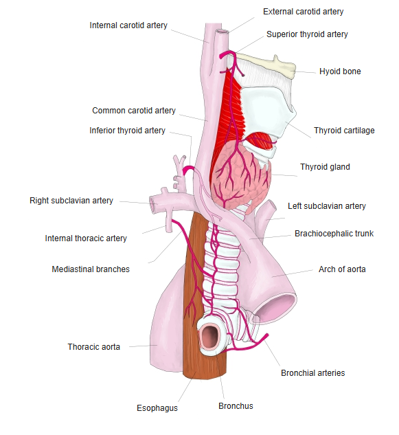
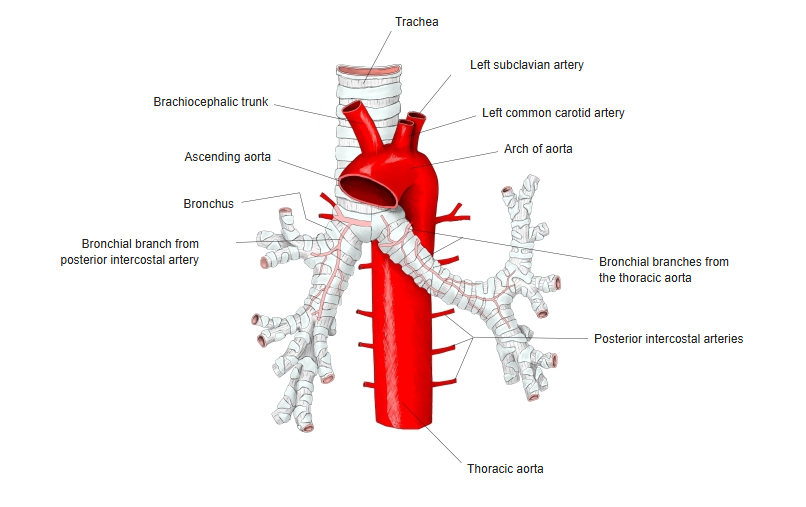
| Arteries | · Bronchial arteries (visceral branches of the thoracic aorta). · Tracheal branches of the inferior thyroid artery, near the bifurcation. · Bronchial branches of the internal thoracic artery. |
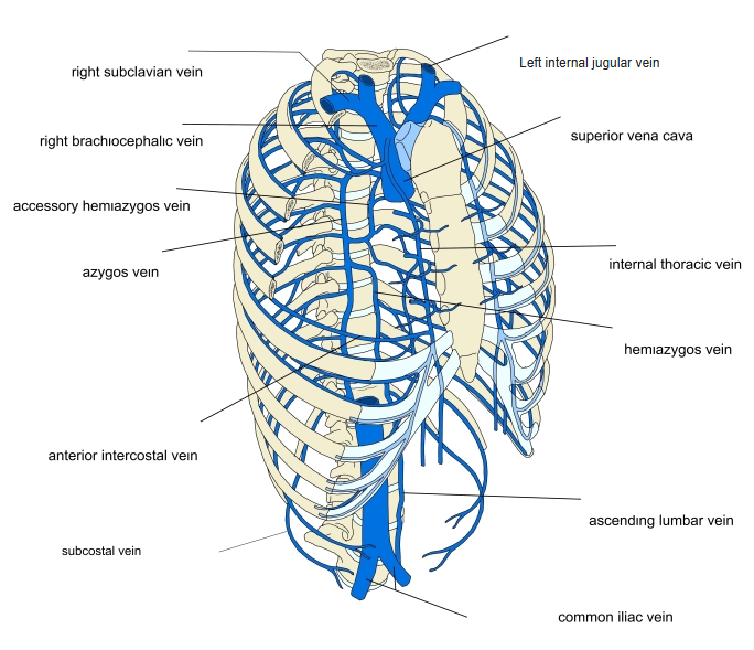
| Veins | Anterior bronchial veins to the brachiocephalic veins; posterior bronchial veins to the azygos vein and the accessory hemiazygos vein. |
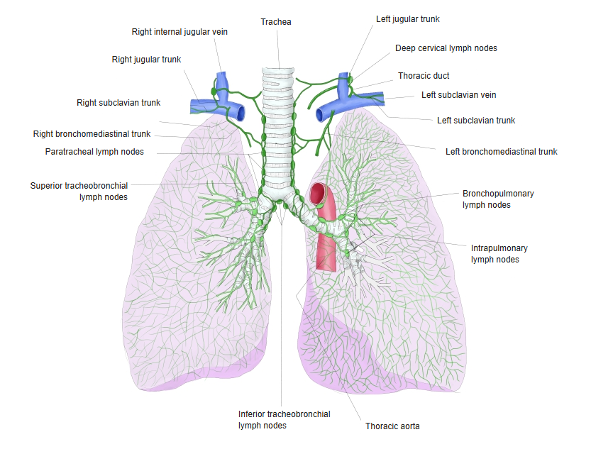
| Lymphatics | Drain to the pretracheal and paratracheal lymph nodes and the superior and inferior tracheobronchial nodes. |
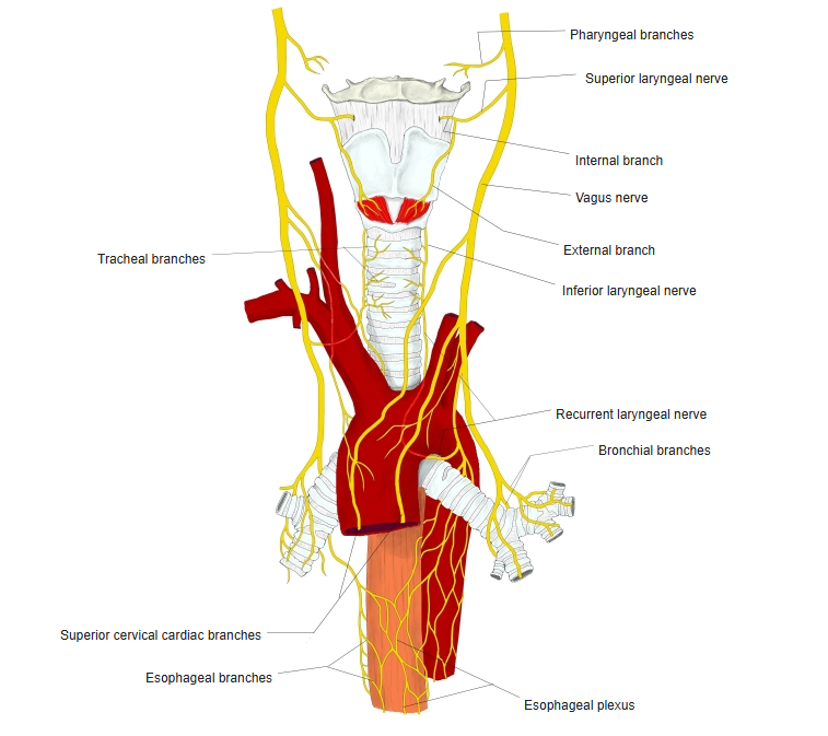
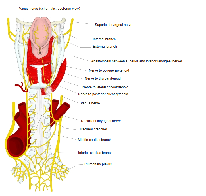
| Nerves | Bronchial branches of the recurrent laryngeal nerve, anterior and posterior bronchial branches of the vagus nerve, and sympathetic branches, joining in the pulmonary plexus. |
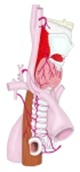 Arteries 1
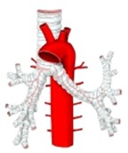 Arteries 2
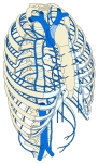 Veins
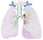 Lymph nodes and lymph trunks
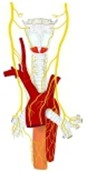 Nerves 1
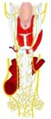 Nerves 2
References and sources
The trachea divides at the carina, at about the level of the sternal angle, into the two main bronchi. The right main bronchus is wider, shorter and more vertical; the azygos vein arches over it, and about 2 cm from its origin it gives off the superior lobar bronchus, which lies above the right pulmonary artery (the eparterial bronchus). The left main bronchus, about 5 cm long, passes below the aortic arch and in front of the esophagus, the thoracic duct and the descending aorta, and has no eparterial branch.
Down the bronchial tree the amount of hyaline cartilage decreases until it is absent in the bronchioles, while the smooth muscle increases. The lining changes from pseudostratified ciliated columnar epithelium to simple cuboidal epithelium and, in the alveoli, simple squamous epithelium, across which oxygen and carbon dioxide are exchanged with the blood in the capillaries.
Variants: in about 0.1–5% of people a right upper lobe bronchus arises from the trachea above the carina (tracheal bronchus); it is usually harmless but can cause recurrent infection. An accessory cardiac bronchus arising from the intermediate bronchus is found in about 0.3%.
- Further reading: Bronchus, Wikipedia.