Latin: musculus frontalis (frontal belly of the occipitofrontalis)
Overview
- Often treated, with the occipitalis, as one muscle: the occipitofrontalis (frontal belly and occipital belly). Both pass into the galea aponeurotica.
- Thin, quadrilateral, closely attached to the superficial fascia; broader than the occipitalis, with longer, paler fibres.
- Has no bony attachment.
- Medial fibres continue from the procerus; intermediate fibres blend with the corrugator supercilii and orbicularis oculi; lateral fibres meet the orbicularis oculi over the zygomatic process of the frontal bone.
- Paired or unpaired? Some textbooks describe it as unpaired, but others describe left and right muscles that join below and separate above; many illustrations show them separately.
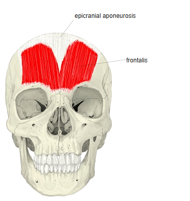
Position in the classification
Head › Facial muscles › Epicranius
- Frontalis
- Occipitalis
- Temporoparietalis
Illustrations
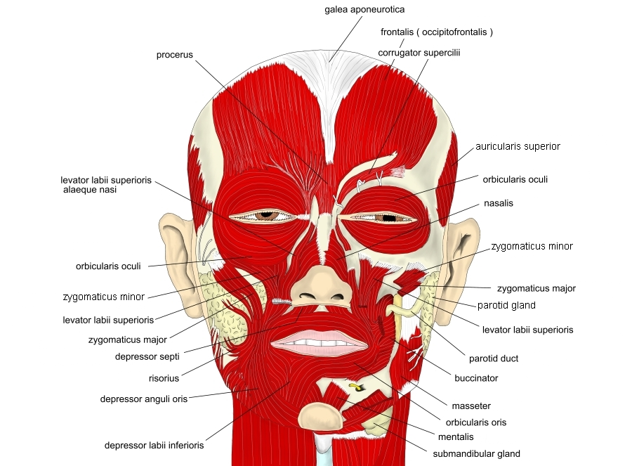
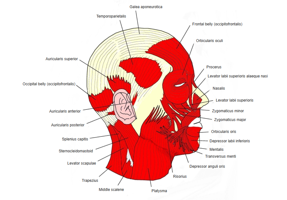
Origin / insertion
- Origin: skin of the root of the nose, the medial angle of the eye and the eyebrows
- Insertion: galea aponeurotica (in front of the coronal suture)
Action
- Raises the eyebrows and makes horizontal wrinkles in the forehead
- Pulls the galea aponeurotica and scalp forward
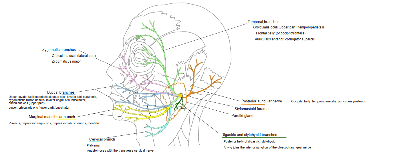
Innervation
- Facial nerve, temporal branches
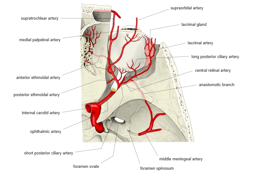
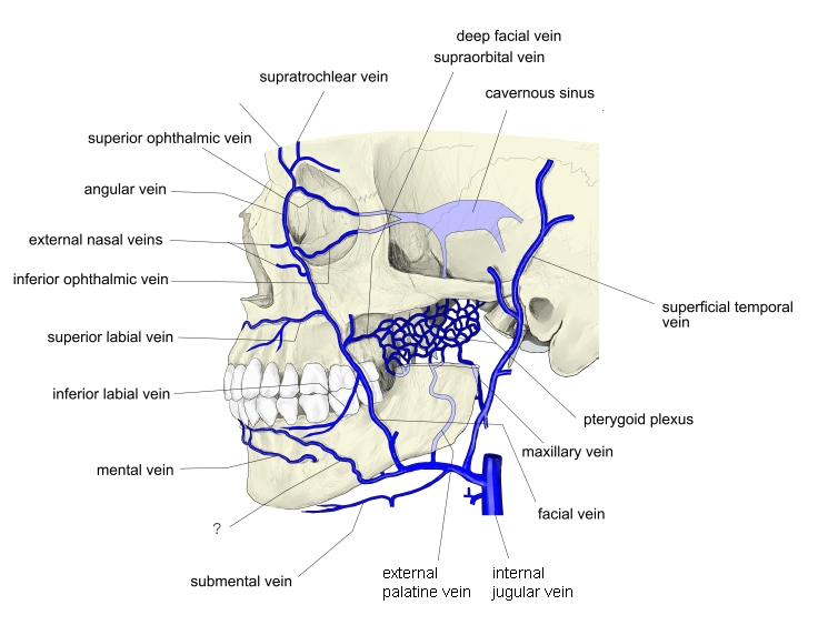
Blood supply
- Supraorbital and supratrochlear arteries (branches of the ophthalmic artery)
- Veins: not described in the sources used here
Anatomical variations
- May be absent
- Fibres of both sides may cross in the midline
- Often divided into several bundles
- Very rarely continuous with the occipitalis
Related terms
- Occipitofrontalis
- Occipitalis
- Galea aponeurotica
- Procerus
- Corrugator supercilii
- Orbicularis oculi
- Facial nerve
- Supraorbital artery
References and sources
- Further reading: Frontalis muscle, Wikipedia.