Right lamina (Latin: lamina dextra cartilaginis thyroideae)
Also called the right lamina of thyroid cartilage
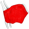 Right lamina (right side)
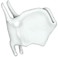 Features of the right lamina
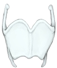 Thyroid cartilage (anterior)
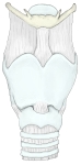 Larynx (anterior)
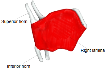
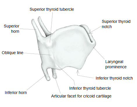
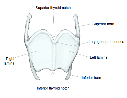
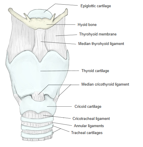
· The right and left laminae are short plates; on each, four borders and two surfaces can be distinguished. In front the two laminae meet at an angle, forming the laryngeal prominence (Adam's apple); behind, they stay apart.
Anterior border: short; it joins the other lamina in the midline.
Posterior border: thick and long, projecting upwards and downwards; the upper projection is the superior horn and the lower one the inferior horn. The inferior horn has a facet for the cricoid cartilage (cricothyroid joint).
Superior border: has the deep superior thyroid notch in the midline.
Inferior border: has the shallow inferior thyroid notch in the midline; the cricothyroid muscle is attached near it.
Internal surface: generally smooth, narrow and slightly concave; near the midline angle it gives attachment to the vocal folds (the vocal ligaments).
External surface: crossed by the oblique line, which runs from behind and above to the front and below, from the superior thyroid tubercle to the inferior thyroid tubercle. Muscles such as the sternothyroid attach here.
Near the superior thyroid tubercle there is often a small thyroid foramen, which can carry the superior laryngeal artery, vein and nerve; it is a variation.
Note: some textbooks call it the foramen of the thyroid cartilage.
Reference websites
[Wikipedia]
· Thyroid cartilage, Wikipedia.
[Websites with illustrations]
· Larynx, external view (labelled diagram) (Wikimedia Commons)

Related terms
- Laryngeal prominence
- Larynx
- Laryngeal cartilages (anterior view)
- Vocal fold
- Sternothyroid
- Cricothyroid
- Superior laryngeal artery
References and sources
- Further reading: Thyroid cartilage, Wikipedia.
- Federative International Programme for Anatomical Terminology (FIPAT). Terminologia Anatomica, 2nd ed. (TA2), 2019.