Canine fossa (Latin: fossa canina)
In more detail:
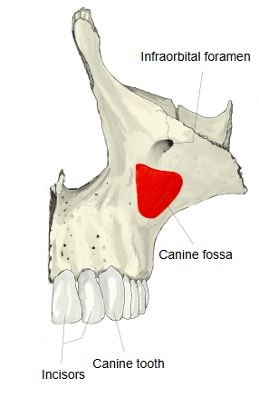
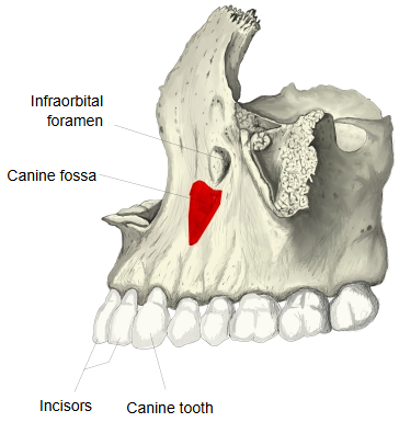
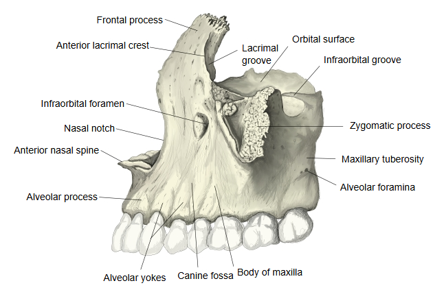
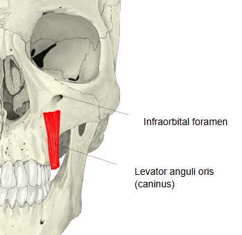
The canine fossa lies on the front of the maxilla between the tooth sockets of the alveolar process and the infraorbital margin, stretching roughly from above the canine to above the first molar. It lies lateral to the incisive fossa, separated from it by the canine eminence, the ridge over the root of the canine tooth. The levator anguli oris arises here, which is why the muscle is also called caninus. The fossa is well marked in modern humans and faint or absent in earlier hominins, and its depth varies between populations; it is thought to reflect a smaller, less projecting maxilla as the chewing apparatus became reduced.
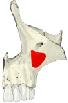 Canine fossa (anterolateral view)
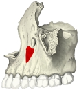 Canine fossa (lateral view)
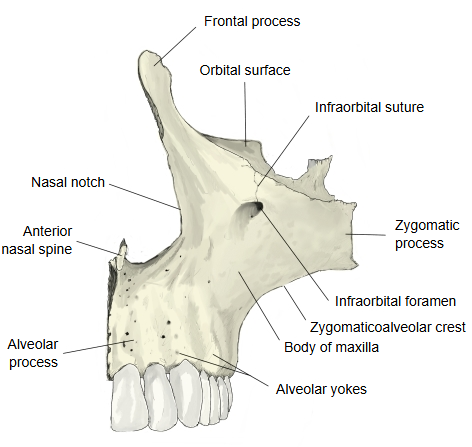
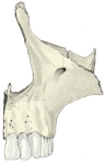 Maxilla (anterior view)
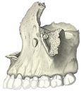 Maxilla (lateral view)
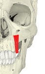 Levator anguli oris
Reference websites
[Wikipedia]
· Canine fossa, Wikipedia.
[Websites with illustrations]
· The maxilla (TeachMeAnatomy)
· Left maxilla, outer surface (Gray's Anatomy) (Wikimedia Commons)

Related terms
- Maxilla
- Body of maxilla
- Infraorbital foramen
- Infraorbital margin
- Alveolar process
- Levator anguli oris
References and sources
- Further reading: Canine fossa, Wikipedia.
- Federative International Programme for Anatomical Terminology (FIPAT). Terminologia Anatomica, 2nd ed. (TA2), 2019.