Body of maxilla (Latin: corpus maxillae)
- Overview
- The four surfaces (anterior, infratemporal, nasal, orbital)
- Reference websites
- (Wikipedia)
- Where it is found
- Related terms
Overview
The body of the maxilla is the main part of the maxilla, what remains when its four processes are taken away; it has four surfaces.
Maxilla: one of the bones of the facial skeleton (viscerocranium)Four processes: frontal, zygomatic, palatine and alveolar processesFour surfaces: anterior, posterior (infratemporal), superior (orbital) and medial (nasal)
· Shape: roughly pyramidal (IMAIOS).
The main feature of the body of the maxilla is the maxillary sinus inside it, which communicates with the nasal cavity through the maxillary hiatus.
Maxillary sinus: the largest of the paranasal sinusesMaxillary hiatus: in the articulated skull it is largely closed by the ethmoid bone, the palatine bone and the inferior nasal concha
- Body of maxilla : the main, roughly pyramid-shaped part of the maxilla; it contains the maxillary sinus and has four surfaces.
- Processes
- Frontal process : a long, flat process reaching upwards, between the nasal bone and the lacrimal bone.
- Zygomatic process : a thick, short process from the lateral side of the body that joins the zygomatic bone.
- Palatine process : projects almost horizontally inwards from the lower part of the body.
- Alveolar process : the downward continuation of the body, holding the sockets for the roots of the teeth.
The four surfaces (anterior, infratemporal, nasal, orbital)
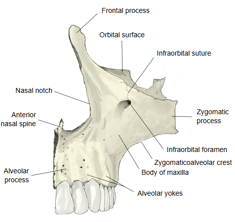
| 1 | Anterior surface | The anterior surface faces forwards and slightly outwards. Above, it reaches up from the frontal process to form part of the infraorbital margin; below the margin opens the infraorbital foramen, which transmits the infraorbital nerve and vessels. Laterally it runs towards the zygomatic process and the zygomaticomaxillary suture. Below the foramen is a shallow hollow, the canine fossa, where levator anguli oris arises; above the incisor teeth is the smaller incisive fossa. Below, the surface continues into the alveolar process. Medially it forms the nasal notch; in the midline the two maxillae meet at the intermaxillary suture, and at the lower end of the notch the anterior nasal spine projects forwards. Laterally it is separated from the infratemporal surface by the zygomaticoalveolar crest. Related terms: infraorbital foramen, infraorbital margin, infraorbital suture, zygomaticomaxillary suture, canine fossa, nasal notch | 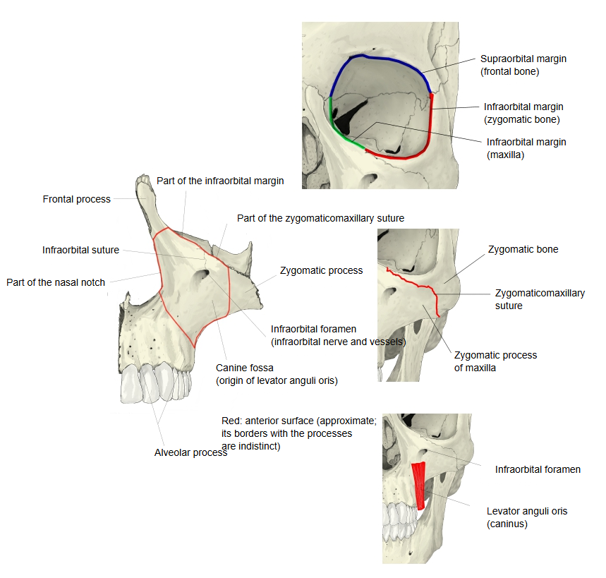 |
| 2 | Infratemporal surface (= posterior surface) | The part below the lower edge of the zygomatic process is the infratemporal surface. It faces backwards and outwards, towards the infratemporal fossa, and is convex; it curves gently from the zygomaticoalveolar crest down to the alveolar process. Towards its back it has two or more small alveolar foramina, which lead into the alveolar canals for the posterior superior alveolar branches of the maxillary nerve and the posterior superior alveolar vessels. Lower down, towards the alveolar process, it forms the rounded, rough maxillary tuberosity behind the last molar. Related terms: zygomaticoalveolar crest, alveolar foramina, maxillary tuberosity, alveolar canals | 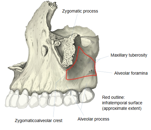 |
| 3 | Nasal surface (= medial surface) | The nasal surface forms part of the lateral wall of the nasal cavity. The large maxillary hiatus opens on it; in the articulated skull it is largely closed by the palatine bone, the inferior nasal concha and other bones. It is the outlet of the maxillary sinus into the nasal cavity. In front of the hiatus the vertical lacrimal groove forms the lateral wall of the nasolacrimal canal; the edge behind the groove meets the lacrimal bone as the lacrimal margin. Small hollows above and behind the hiatus meet the ethmoidal labyrinth and the orbital process of the palatine bone. Behind the hiatus a rough area meets the perpendicular plate of the palatine bone; across its lower back part the greater palatine groove runs obliquely down and, with the matching groove of the palatine bone, forms the greater palatine canal for the greater palatine nerve and vessels. Lower in front, the conchal crest articulates with the inferior nasal concha. Related terms: maxillary hiatus, lacrimal groove, lacrimal margin, greater palatine groove | 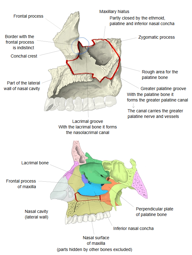 |
| 4 | Orbital surface (= superior surface) | The orbital surface is smooth and triangular and forms most of the floor of the orbit. The infraorbital groove begins near the middle of its posterior border and runs obliquely forwards; it becomes the infraorbital canal, which opens at the infraorbital foramen on the anterior surface. Running from the canal to the infraorbital margin is the infraorbital suture, the line where the roof of the groove closed over to form the canal. The orbital surface has three borders: • Anterior border: part of the infraorbital margin. • Posterior border: the lower edge of the inferior orbital fissure. • Medial border: meets the lacrimal bone, the orbital plate of the ethmoid bone and the orbital process of the palatine bone. Related terms: infraorbital groove, infraorbital suture | 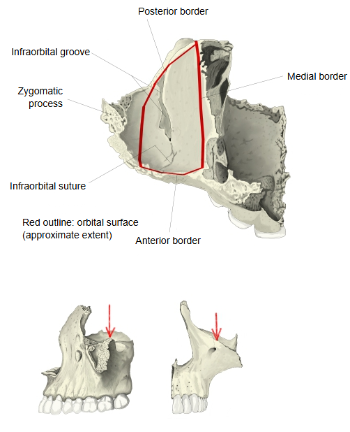 |
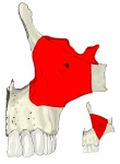 Body of maxilla (anterior)
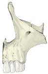 Right maxilla (anterolateral)
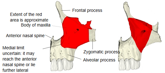
Reference websites
[Wikipedia]
· Body of maxilla, Wikipedia.
Anatomy reference sites describe it as follows.
The body is roughly pyramidal and contains a large cavity, the maxillary sinus (antrum of Highmore). It has four surfaces: anterior, posterior or infratemporal, superior or orbital, and medial or nasal. Where the surfaces meet the processes the borders are not sharply defined, so the extent of each surface varies between sources.
Where it is found
Related terms
- Maxillary sinus
- Maxillary hiatus
- Frontal process
- Zygomatic process
- Alveolar process
- Palatine process
- Infraorbital foramen
References and sources
- Further reading: Body of maxilla, Wikipedia.
- Further reading: Maxilla, Wikipedia.
- Federative International Programme for Anatomical Terminology (FIPAT). Terminologia Anatomica, 2nd ed. (TA2), 2019.