Infraorbital foramen (Latin: foramen infraorbitale)
Also called the infra-orbital foramen
Its position and its place in the course of the nerve can be described as follows.
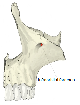
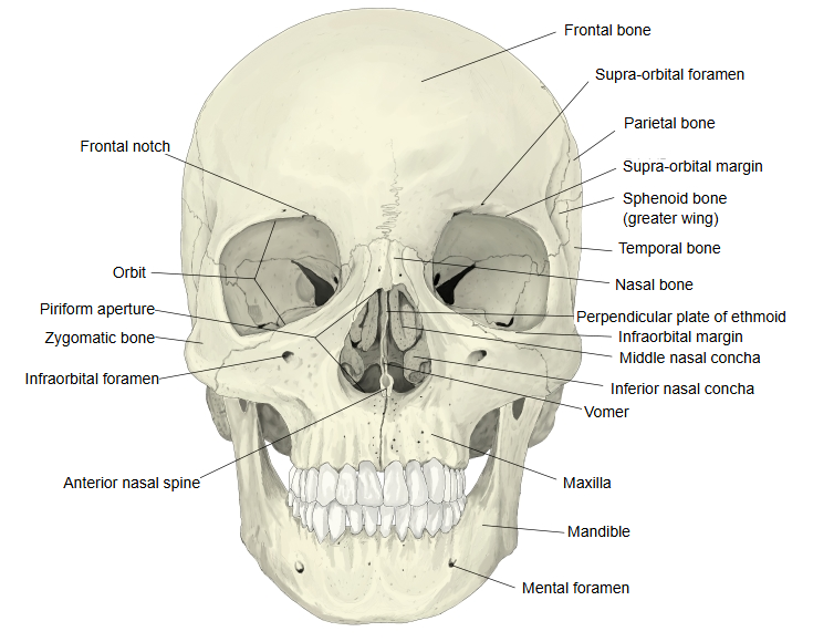
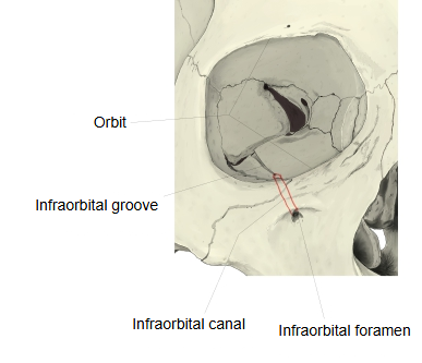
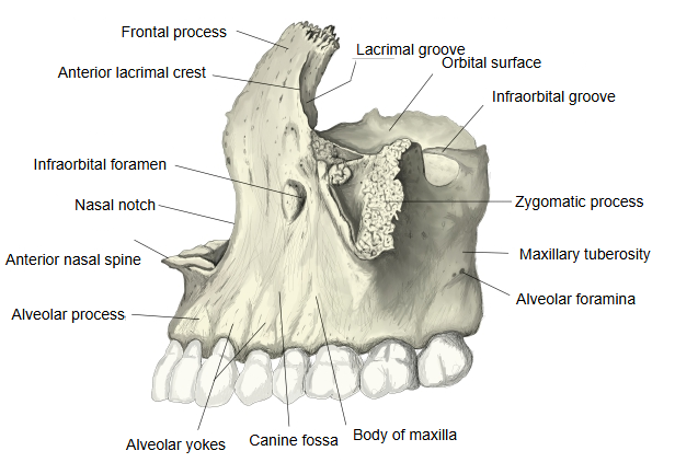
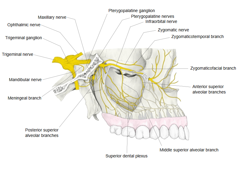
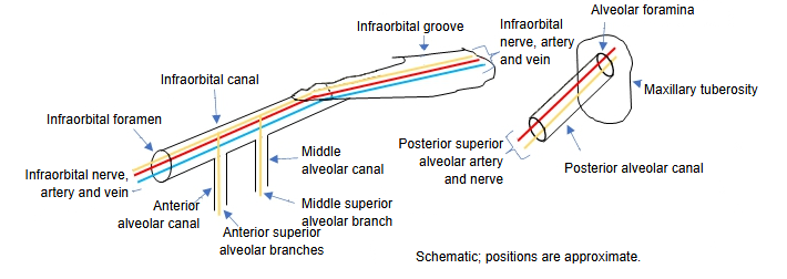
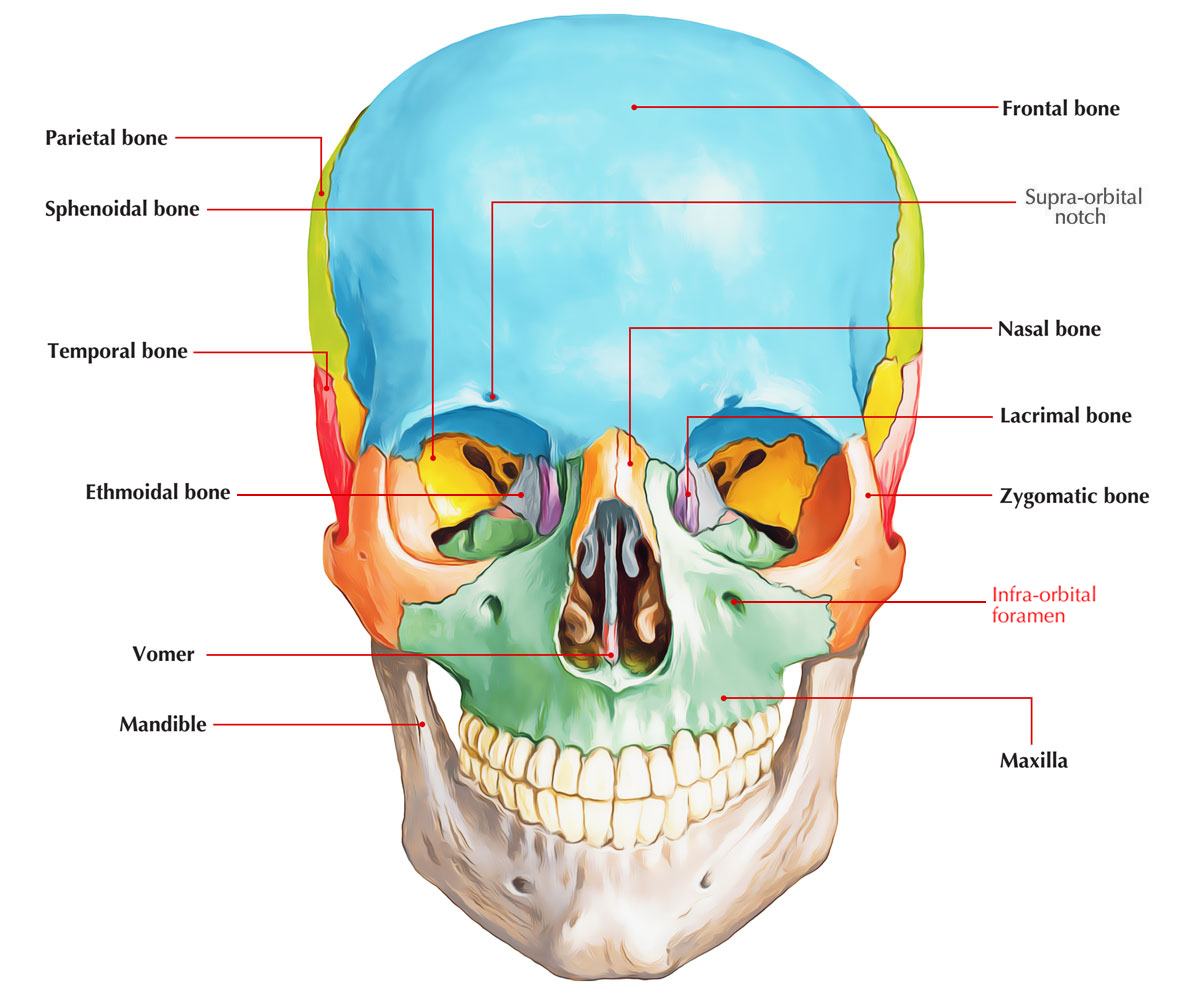
The lower border of the body of the maxilla passes without a clear boundary into the alveolar process. About 0.5-1 cm below the infra-orbital margin lies the infraorbital foramen (sources differ slightly; most give roughly 6-11 mm), the opening on the face of the infraorbital canal, which continues the infraorbital groove of the orbital surface. The nerve it carries is the end of the maxillary nerve. The supra-orbital, infraorbital and mental foramina lie roughly on one vertical line through the pupil. The foramen is the usual site for an infraorbital nerve block, and pressing over it can be used to test the nerve. Branches of the canal, the alveolar canals, carry the superior alveolar nerves and vessels down to the upper teeth.
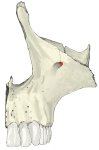 Maxilla (anterolateral view)
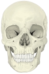 Skull (anterior view)
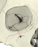 Infraorbital groove, canal and foramen
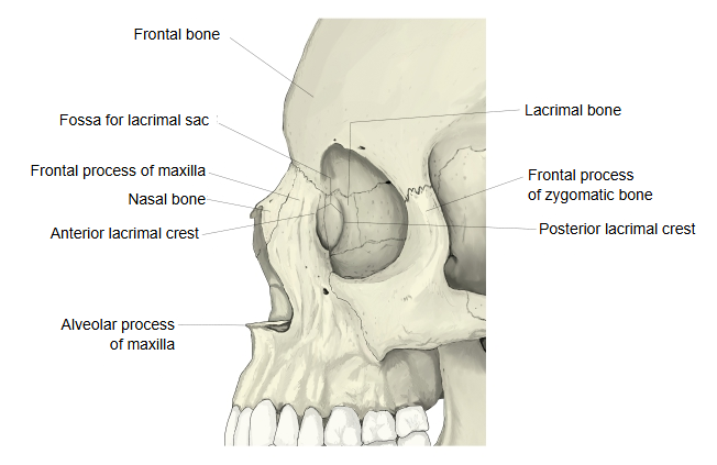
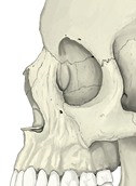 Orbit and maxilla (anterolateral view)
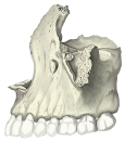 Maxilla (lateral view)
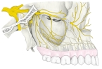 Maxillary nerve and its branches
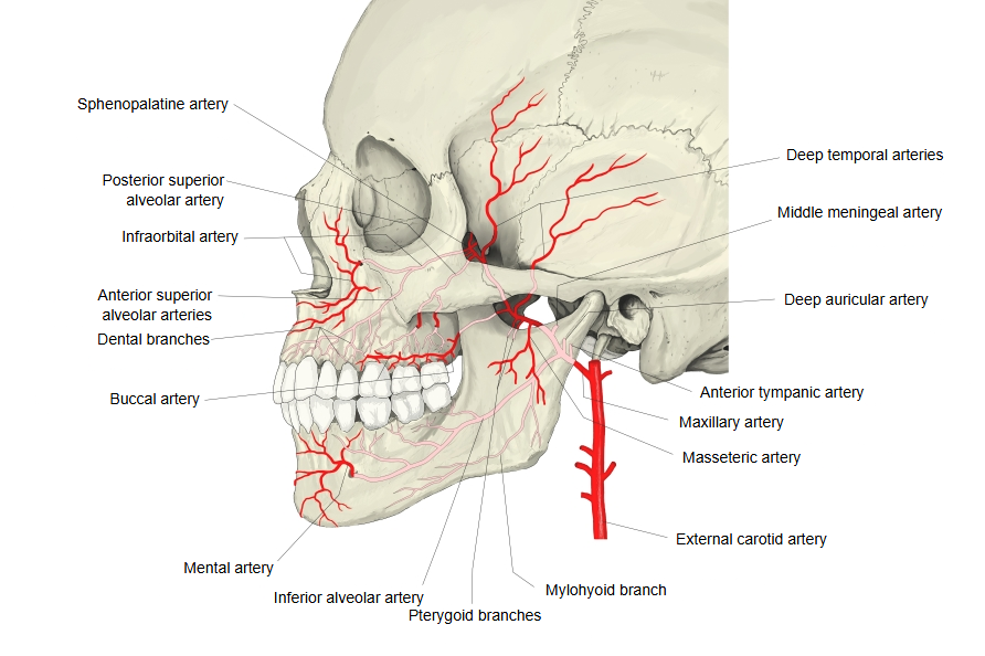
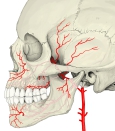 Maxillary artery and its branches
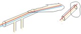 Infraorbital and alveolar canals (diagram)
Reference websites
[Wikipedia]
· Infraorbital foramen, Wikipedia.
· Infraorbital nerve, Wikipedia.
[Websites with illustrations]
· Infraorbital foramen (skull, anterior view) (Wikimedia Commons)

· Infraorbital foramen (close-up) (Wikimedia Commons)

· Infraorbital foramen (Earth's Lab)
Where it is found
Related terms
References and sources
- Further reading: Infraorbital foramen, Wikipedia.
- Further reading: Infraorbital nerve, Wikipedia.
- Federative International Programme for Anatomical Terminology (FIPAT). Terminologia Anatomica, 2nd ed. (TA2), 2019.