Latin: musculi lumbricales pedis
Overview
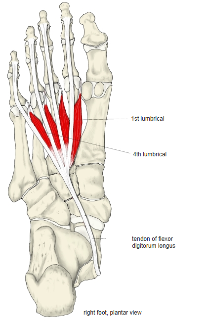
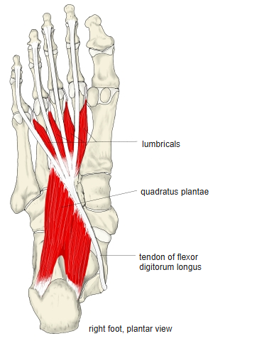
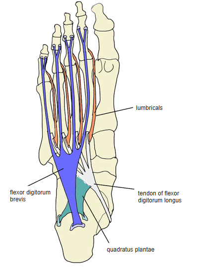
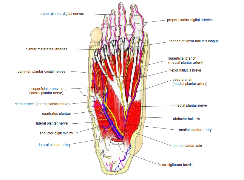
- Four small muscles attached to the tendons of the flexor digitorum longus, numbered from the medial side.
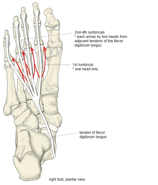
- The 1st has one head; the 2nd–4th have two heads from the adjacent tendons.
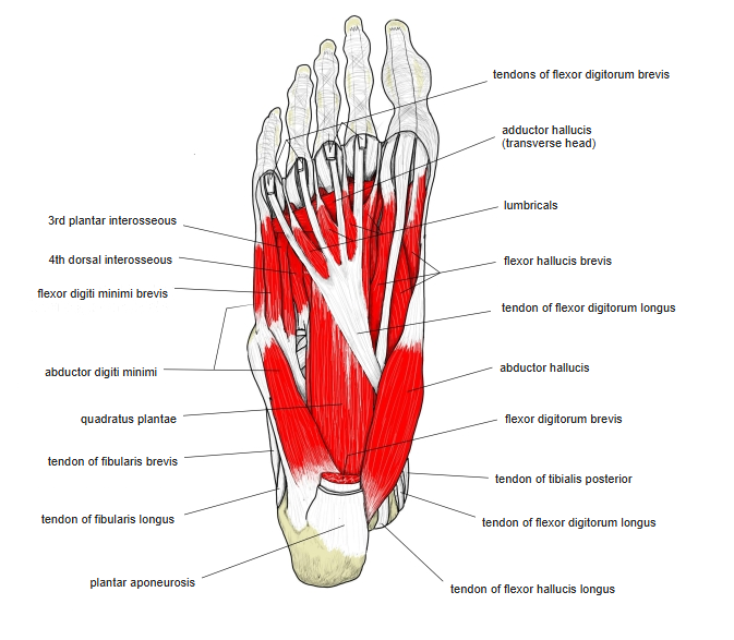
- They run on the plantar side of the deep transverse metatarsal ligament, separated from it by small bursae.
- One or more may be absent or doubled; they cannot be palpated.
Position in the classification
Lower Limb › Foot › Sole of foot › Metatarsal muscles
Illustrations
Origin / insertion
- Origin: the divided tendons of the flexor digitorum longus (1st: one head; 2nd–4th: two heads)
- Insertion: dorsal (extensor) expansions of the 2nd–5th toes, passing along their medial (big-toe) side, over the proximal phalanges
Action
- Flex the proximal phalanges of the 2nd–5th toes (MTP joints)
- Some sources add: extension of the PIP and DIP joints and drawing the toes towards the big toe
- Help to prevent the toes from curling (clawing) when the flexor digitorum longus contracts
Innervation
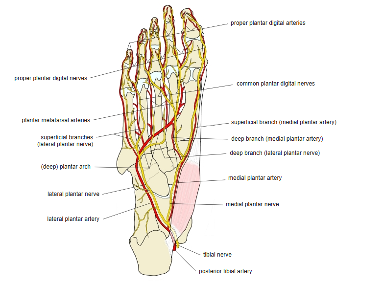
- Two nerves (sources differ):
- 1st: medial plantar nerve; 2nd–4th: deep branch of the lateral plantar nerve
- Some sources: 1st and 2nd by the medial plantar nerve (L5, S1), 3rd and 4th by the lateral plantar nerve (S1, S2)
- There is much individual variation
Blood supply
- Medial and lateral plantar arteries
Anatomical variations
- One or more may be absent; the 3rd or 4th may be doubled; the insertion may be partly or wholly into the proximal phalanges
Related terms
- Flexor digitorum longus
- Quadratus plantae
- Lumbricals (hand)
- Medial plantar nerve
- Lateral plantar nerve
References and sources
- Further reading: Lumbricals of the foot, Wikipedia.
- Gray H. Anatomy of the Human Body, 20th ed. (1918), “8e. The Muscles and Fasciæ of the Foot”, Bartleby.com.