Membranous ampullae (Latin: ampullae membranaceae)
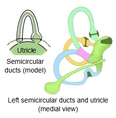
· Each semicircular duct has one, so there are three: the anterior, the lateral and the posterior membranous ampulla.
- Anterior semicircular duct : the uppermost. Of its two limbs, the ampullary limb forms the anterior membranous ampulla just before it opens into the utricle; the simple limb joins that of the posterior duct to form the membranous common crus.
- Lateral semicircular duct : the most lateral. Its ampullary limb forms the lateral membranous ampulla just before it opens into the utricle; its simple limb opens into the utricle on its own.
- Posterior semicircular duct : the lowest. Its ampullary limb forms the posterior membranous ampulla just before it opens into the utricle; its simple limb joins that of the anterior duct to form the membranous common crus.
The ducts are filled with endolymph and are fixed to the walls of the bony semicircular canals by strands of connective tissue. Each membranous ampulla lies inside the matching bony ampulla, surrounded by perilymph.
- Anterior duct : ampullary limb → anterior membranous ampulla → utricle; simple limb → membranous common crus → utricle.
- Lateral duct : ampullary limb → lateral membranous ampulla → utricle; simple limb → directly into the utricle.
- Posterior duct : ampullary limb → posterior membranous ampulla → utricle; simple limb → membranous common crus (with the anterior duct) → utricle.
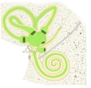 Bony and membranous labyrinth
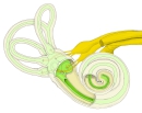 Labyrinth and nerves
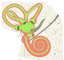 Perilymphatic space and membranous labyrinth
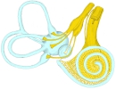 Membranous labyrinth and nerves
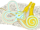 Labyrinth and nerves (detail)
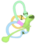 The three semicircular ducts
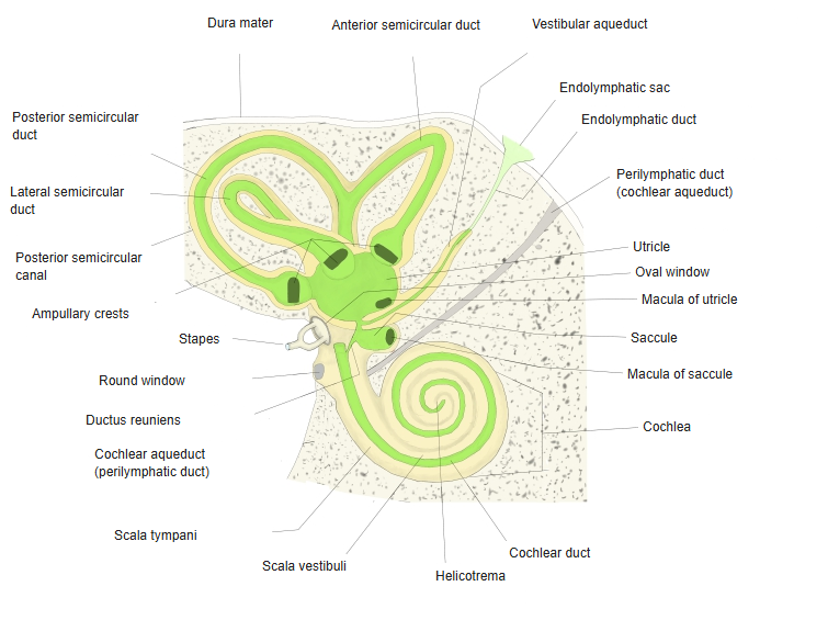
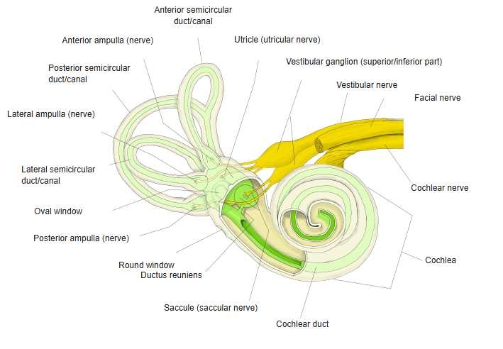
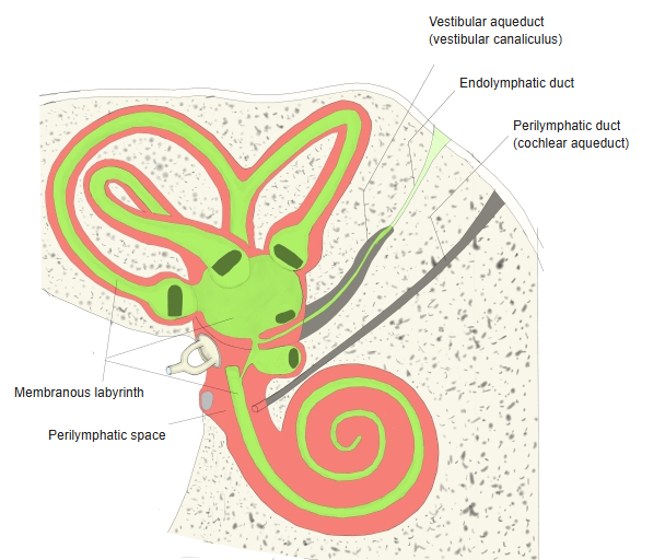
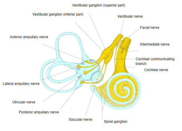
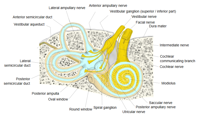
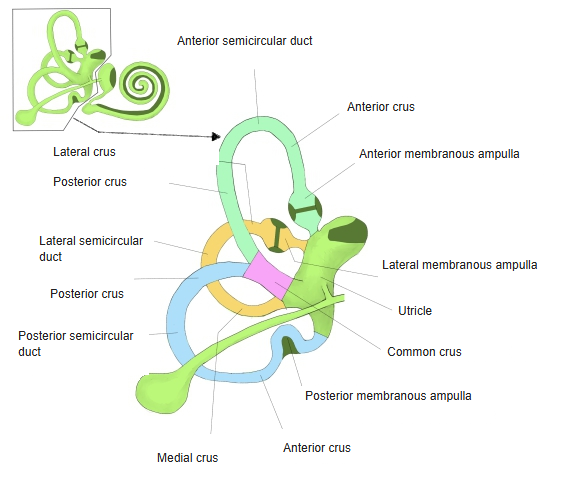
The ampullae and ampullary crests of the anterior and lateral ducts lie very close together, whereas those of the posterior duct lie far from them, almost on the opposite side. The three ampullae have about the same shape and size: about 2–2.5 mm in the direction of the duct and 1.5 mm across it. The nerve enters on the convex side of the duct, marked by a single transverse groove, the ampullary groove. The ampullary crest is crescent-shaped, runs across the ampulla and occupies one third to one half of its circumference.
The ampullary crest carries hair cells whose hair bundles project into a gelatinous cap, the cupula. Flow of the endolymph when the head rotates bends the cupula and stimulates the hair cells; the signals travel in the ampullary nerves of the vestibular nerve.
Reference websites
[Wikipedia]
· Semicircular canals, Wikipedia.
· Crista ampullaris, Wikipedia.
[Websites with illustrations]
· The inner ear: bony labyrinth and membranous labyrinth (TeachMeAnatomy)
· Internal ear anatomy: cochlea, vestibule and canals (Kenhub)
Related terms
- Semicircular ducts
- Membranous labyrinth
- Utricle
- Ampullary crest
- Ampullary groove
- Lateral membranous ampulla
- Posterior membranous ampulla
References and sources
- Further reading: Semicircular canals, Wikipedia.
- Further reading: Crista ampullaris, Wikipedia.
- Federative International Programme for Anatomical Terminology (FIPAT). Terminologia Anatomica, 2nd ed. (TA2), 2019.