Raschkow plexus
Also called the plexus of Raschkow or the subodontoblastic nerve plexus or the parietal layer of nerves
of the
pulp
- Odontoblastic layer : the outermost zone: a single layer of odontoblasts along the dentin, sending long processes (dentinal fibres) into the dentin. Rich in blood vessels.
- Cell-free zone : few cells, but rich in capillaries and nerve fibres; often hard to see in the root pulp.
- Cell-rich zone : many fibroblasts and undifferentiated mesenchymal cells; rich in nerves: the Raschkow plexus lies here and just inside it.
- Pulp core : the central mass of the pulp inside these three zones, with the larger vessels and nerve bundles.
Vessels and nerves are arranged in the pulp as follows.
Blood vessels are plentiful from the odontoblastic layer to the cell-free zone, and the Raschkow plexus is developed between the cell-free zone and the cell-rich zone. Some branches from the plexus lose their myelin, cross the cell-free zone and enter the inner part of the dentinal tubules beside the processes of the odontoblasts.
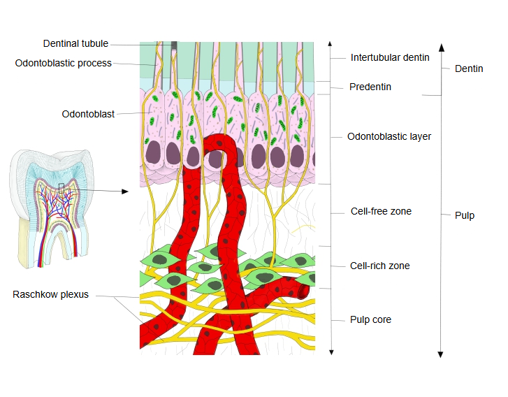
· Nerve bundles enter the pulp through the apical foramen together with the blood vessels, run up through the pulp core and branch toward the periphery; beneath the odontoblastic layer they spread out into the plexus, which is best developed in the crown.
· Most of its fibres are sensory (thin myelinated A-delta and unmyelinated C fibres) from the trigeminal nerve, through the superior or inferior alveolar nerves, and carry pain from the pulp and dentin. Autonomic fibres, mostly sympathetic, mainly control the flow of blood in the pulp.
· The plexus is not fully formed until the root of the tooth is complete. Sources differ on its exact level: it is placed in the cell-rich zone, or between the cell-rich zone and the pulp core.
Reference websites
[Wikipedia]
· Pulp (tooth), Wikipedia.
[Websites with illustrations]
· Pulp (Pocket Dentistry)
· Tooth anatomy (Blausen) (Wikimedia Commons)

Related terms
References and sources
- Further reading: Pulp (tooth), Wikipedia.
- Federative International Programme for Anatomical Terminology (FIPAT). Terminologia Anatomica, 2nd ed. (TA2), 2019.