Spiral ganglion (Latin: ganglion spirale cochleae)
Also called Cochlear ganglion
· It is also called the cochlear ganglion.
'Spiral ganglion' is the usual name; 'cochlear ganglion', named after the organ, is found less often.
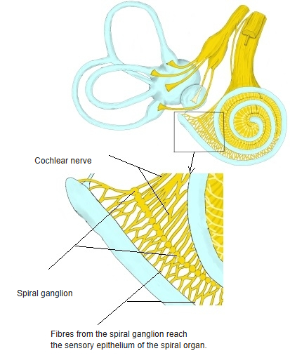
· Its nerve cell bodies lie in the spiral canal of the modiolus, at the root of the osseous spiral lamina, and follow the turns of the cochlea up towards the cochlear cupula (apex) like a string of beads.
· It contains roughly 30,000–50,000 neurons; sources differ on the exact number.
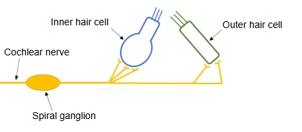
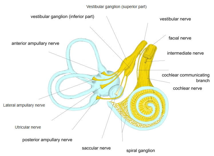
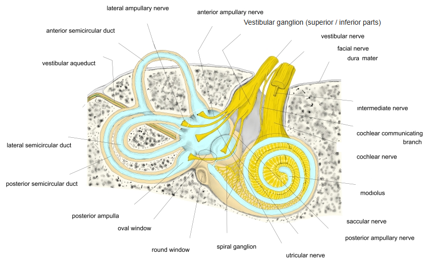
· The cells are bipolar. Their peripheral processes run out through the osseous spiral lamina to the sensory epithelium of the spiral organ (organ of Corti), the inner and outer hair cells; their central processes gather in the modiolus to form the cochlear nerve. Type I cells (about 90–95%) are large, myelinated and supply the inner hair cells. Type II cells (the rest) are small, mostly unmyelinated and each supplies many outer hair cells.
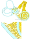 Spiral ganglion in the cochlea
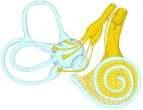 Cochlear and vestibular nerves
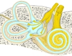 Vestibulocochlear nerve in the inner ear
Reference websites
[Wikipedia]
· Spiral ganglion, Wikipedia.
Related terms
- Spiral limbus
- Spiral prominence
- Hamulus of spiral lamina
- Foraminous spiral tract
- Cochlear nerve
- Vestibulocochlear nerve
- Modiolus
- Spiral canal of modiolus
- Osseous spiral lamina
- Spiral organ (organ of Corti)
- Cochlea
References and sources
- Further reading: Spiral ganglion, Wikipedia.
- Federative International Programme for Anatomical Terminology (FIPAT). Terminologia Anatomica, 2nd ed. (TA2), 2019.