Spiral canal of modiolus (Latin: canalis spiralis modioli)
Also called Rosenthal's canal
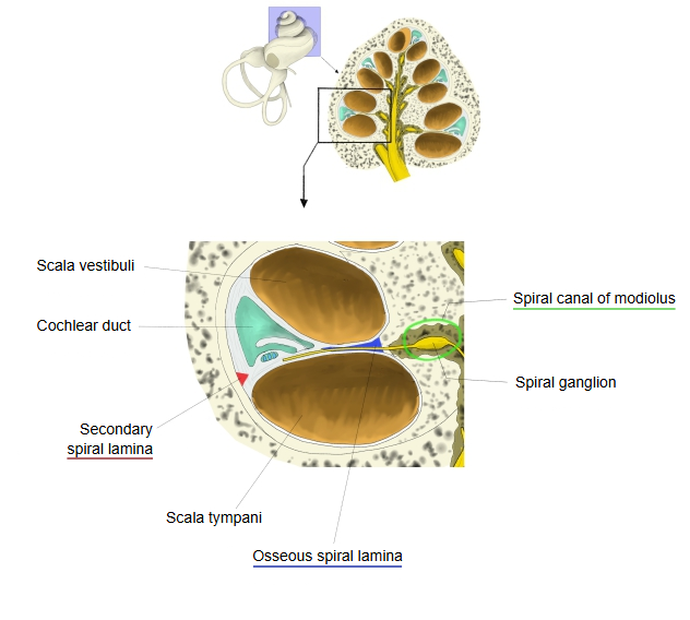
- Spiral canal of the cochlea : total length about 3 cm; 2.5–2.75 turns.
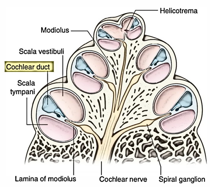
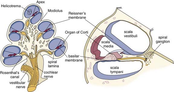
- Upper level: Scala vestibuli : filled with perilymph; passes the sound vibrations to the cochlear duct; part of the bony labyrinth. It joins the scala tympani at the helicotrema.
- Vestibular (Reissner's) membrane : between the scala vestibuli and the cochlear duct.
- Cochlear duct : filled with endolymph; holds the spiral organ; both ends are closed. Contents of the duct region: basilar membrane, organ of Corti, internal and external spiral sulci, spiral prominence, stria vascularis, tectorial membrane.
- Osseous spiral lamina + basilar membrane : (+ secondary spiral lamina) between the cochlear duct and the scala tympani.
- Lower level: Scala tympani : part of the bony labyrinth, filled with perilymph; the travelling wave passes on into it.
- Modiolus : the cone of spongy bone at the centre of the cochlea, with the longitudinal canals and the spiral canal of the modiolus.
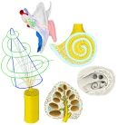 Modiolus (schematic model)
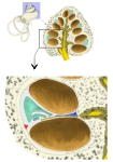 Spiral canal of cochlea (one turn)
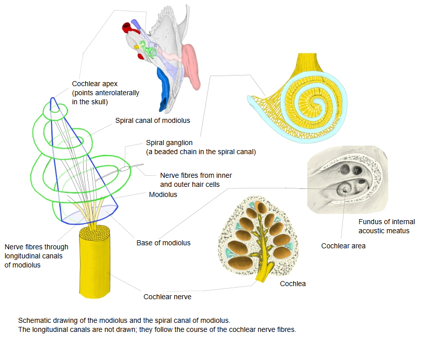
The modiolus is a cone of spongy bone whose base, the base of the modiolus, faces the fundus of the internal acoustic meatus; near the apex, where the second and third turns of the spiral canal of the cochlea wind around it, it becomes a thin plate, the lamina of the modiolus. It contains two kinds of canals. The longitudinal canals carry the fibres of the cochlear nerve and veins up through the modiolus; along the way they send their fibres outward into the spiral canal of the modiolus (see below), and toward the apex the canals themselves end blindly; the large one in the centre is the central canal of the modiolus, and the openings of the others are arranged in a spiral, the foraminous spiral tract. The other canal runs spirally near the edge of the modiolus at the level of the osseous spiral lamina: this is the spiral canal of the modiolus, which holds the spiral ganglion of the cochlear nerve.
The canals of the foraminous spiral tract rise through the modiolus and bend outward one after another toward the attached margin of the osseous spiral lamina. There they widen and run together to form the spiral canal of the modiolus, which follows the attached margin of the lamina and lodges the spiral ganglion: the cell bodies of the cochlear nerve fibres, whose peripheral processes pass through the osseous spiral lamina to the hair cells of the spiral organ.
Reference websites
[Wikipedia]
· Spiral ganglion, Wikipedia.
· Modiolus (cochlea), Wikipedia.
[Websites with illustrations]
· Section of the cochlea (figure) (Earth's Lab)
· Cochlea and modiolus (figure) (Entokey)
Related terms
- Cochlea
- Cochlear nerve
- Spiral organ (organ of Corti)
- Cochlear duct
- Vestibulocochlear nerve
- Internal ear
References and sources
- Further reading: Spiral ganglion, Wikipedia.
- Further reading: Modiolus (cochlea), Wikipedia.
- Federative International Programme for Anatomical Terminology (FIPAT). Terminologia Anatomica, 2nd ed. (TA2), 2019.