Median umbilical fold (Latin: plica umbilicalis mediana)
The median umbilical fold is one of the folds seen on the front wall of the peritoneum: a single fold in the midline that joins the umbilicus and the apex of the urinary bladder.
Fold: a part of the peritoneum raised by a vessel or cord running beneath itIt contains the median umbilical ligament (the remnant of the fetal urachus)
· It is one of the folds of the abdominal wall and has no function after birth. If the urachus stays partly open, a cyst, sinus or fistula can form along its course.
· On each side, the supravesical fossa lies between the median and the medial umbilical fold, above the bladder. Together with the paired medial and lateral umbilical folds it makes five folds on the lower anterior abdominal wall.
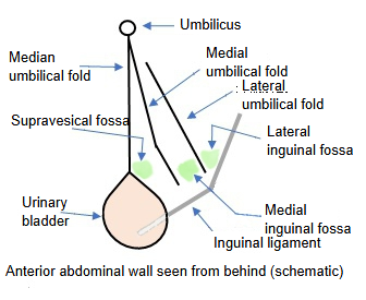
Folds and fossae of the anterior wall of the peritoneum
- Median umbilical fold : in the midline; joins the umbilicus and the apex of the bladder. It contains the median umbilical ligament.
- Supravesical fossa : between the lower parts of the median and medial umbilical folds.
- Medial umbilical fold : paired, lateral to the median umbilical fold. It contains the cord of the umbilical artery.
- Medial inguinal fossa : between the lower parts of the medial and lateral umbilical folds.
- Lateral umbilical fold : paired, lateral to the medial umbilical fold. It contains the inferior epigastric artery and vein.
- Lateral inguinal fossa : between the lower part of the lateral umbilical fold and the inguinal ligament, at the entrance of the inguinal canal (deep inguinal ring).
- Inguinal ligament : the band stretched between the pubic tubercle and the anterior superior iliac spine.
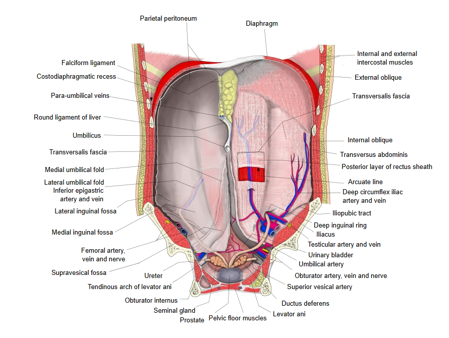
- Fold of the ductus deferens / round ligament of the uterus : formed by the ductus deferens (or the round ligament of the uterus), which passes from the deep inguinal ring, at the lower end of the lateral umbilical fold, medially and backwards into the pelvic cavity.
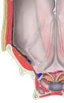 Folds and fossae of the anterior abdominal wall (abdominal cavity seen from behind)
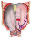 Anterior abdominal wall (abdominal cavity seen from behind)
Reference websites
[Wikipedia]
· Median umbilical fold, Wikipedia.
· Urachus, Wikipedia.
[Websites with illustrations]
· Posterior surface of the anterior abdominal wall: umbilical folds and fossae (figure) (Memorang)

· Median, medial and lateral umbilical ligaments (video thumbnail) (YouTube)

Related terms
- Ventral mesogastrium
- Median umbilical ligament
- Medial umbilical fold
- Lateral umbilical fold
- Supravesical fossa
- Peritoneal folds
- Urinary bladder
- Parietal peritoneum
References and sources
- Further reading: Median umbilical fold, Wikipedia.
- Further reading: Urachus, Wikipedia.
- Federative International Programme for Anatomical Terminology (FIPAT). Terminologia Anatomica, 2nd ed. (TA2), 2019.