Common tendinous ring (Latin: anulus tendineus communis)
Also called: annulus of Zinn
Common tendinous ring
Note: the list aims to be complete, but some structures may have been missed, and sources differ on a few of them (see below).
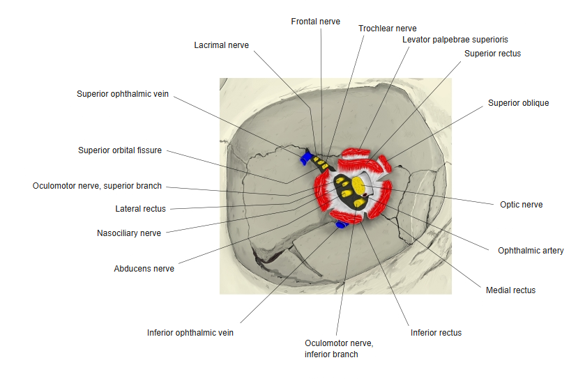
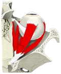 Right orbit (superior view)
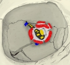 Apex of right orbit
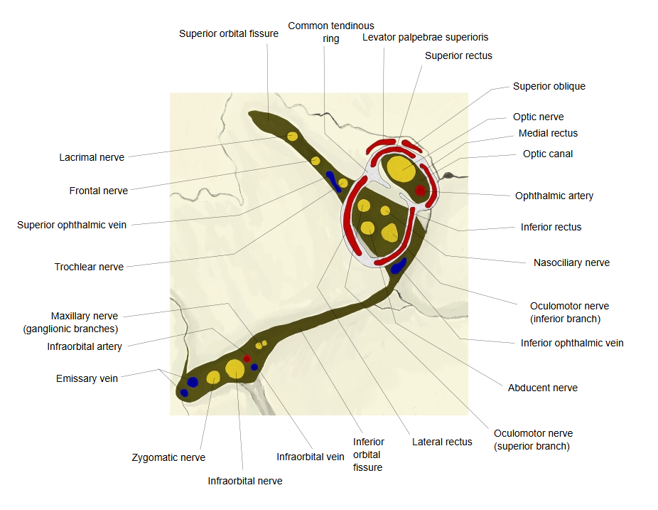
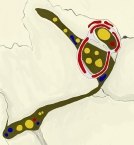 Right orbital fissures
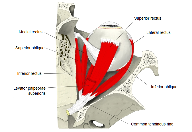
Medical dictionaries describe it as follows.
It is a thickening of the periosteum of the orbit (the periorbita) at the back of the bony orbit, around the optic canal and the middle part of the superior orbital fissure. The four rectus muscles take origin from it, so they form a cone that opens forwards around the eyeball.
Inside the ring: through the optic canal pass the optic nerve and the ophthalmic artery; through the superior orbital fissure pass the superior and inferior branches of the oculomotor nerve, the nasociliary nerve and the abducens nerve. The inferior ophthalmic vein passes through the ring in some descriptions and below it in others.
Outside the ring: above it, through the lateral part of the superior orbital fissure, pass the lacrimal, frontal and trochlear nerves and the superior ophthalmic vein; these therefore lie outside the cone of the rectus muscles. The levator palpebrae superioris and the superior oblique arise from the bone just above and medial to the ring, not from the ring itself; sources differ on how much of the superior oblique is attached to it.
Reference websites
[Wikipedia]
· Annulus of Zinn, Wikipedia.
· Common tendinous ring, The Free Dictionary (medical dictionary).
[Websites with illustrations]
· Muscles of the orbit and the tendinous ring (tryphonov.ru)

· Apex of the orbit with the common tendinous ring (Clinical Gate)

Related terms
- Extra-ocular muscles
- Orbit
- Superior orbital fissure
- Optic canal
- Optic nerve
- Oculomotor nerve
- Abducens nerve
References and sources
- Further reading: Annulus of Zinn, Wikipedia.
- Federative International Programme for Anatomical Terminology (FIPAT). Terminologia Anatomica, 2nd ed. (TA2), 2019.