Arterial grooves (Latin: sulci arteriosi)
Also called the arterial sulci
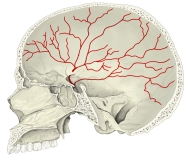 Arterial grooves (median section of skull)
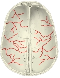 Arterial grooves (calvaria, internal surface)
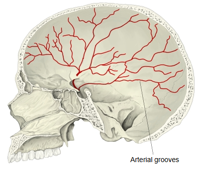
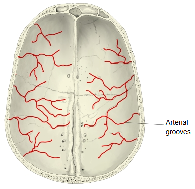
Arterial grooves are the grooves on the inner surface of the skull in which arteries run, between the bone and the dura mater. The grooves of the middle meningeal artery are especially well marked.
· They are most obvious on the inner surface of the parietal bone, the squamous part of the temporal bone and the greater wing of the sphenoid, on the walls of the calvaria and the middle cranial fossa.
· Most are made by the frontal and parietal branches of the middle meningeal artery, which run upwards and backwards from the foramen spinosum. The grooves are deeper near the base and become finer as the branches spread over the vault.
· Because the vessels lie in the bone grooves, a skull fracture here can tear them and cause an extradural (epidural) haemorrhage.
Related terms
- Middle meningeal artery
- Calvaria
- Cranial cavity
- Parietal bone
- Groove for middle temporal artery
- Groove for occipital artery
- Groove for superior sagittal sinus
References and sources
- Further reading: Middle meningeal artery, Wikipedia.
- Further reading: Parietal bone, Wikipedia.
- Federative International Programme for Anatomical Terminology (FIPAT). Terminologia Anatomica, 2nd ed. (TA2), 2019.