Greater wing (Latin: ala major ossis sphenoidalis)
Also called the greater wing of sphenoid
- Surfaces
- Margins
- Openings
- Reference websites
- (Wikipedia, websites with illustrations)
- Where it is found
- Related terms
bone
- Body : the central part of the sphenoid; it contains the sphenoidal sinus and has several surfaces.
- Processes
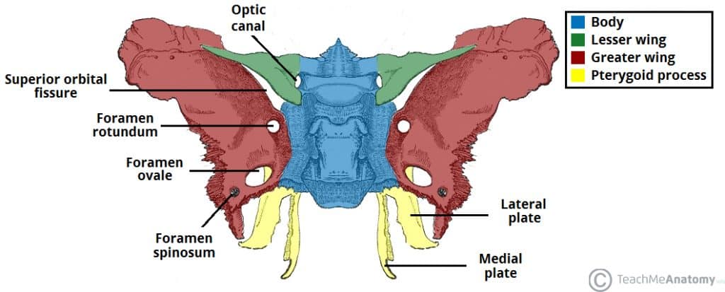
- Greater wing : the largest process; part of the middle cranial fossa and of the side wall of the skull. Three surfaces (cerebral; lateral = temporal and infratemporal; medial = orbital and maxillary) and three margins (upper, anterior, posterior). Openings and features: foramen rotundum, foramen ovale, foramen spinosum, spine of the sphenoid, infratemporal crest.
- Lesser wing : projects to each side from the front of the body; part of the anterior cranial fossa.
- Pterygoid process : projects downwards; made of a medial plate and a lateral plate.
It spreads forwards and laterally like a wing to form part of the middle cranial fossa; its front end is pointed and turns slightly upwards, and forms part of the side wall of the skull. It also forms the back of the lateral wall of the orbit and most of the roof of the infratemporal fossa. Together with the lesser wing it bounds the superior orbital fissure.
Surfaces
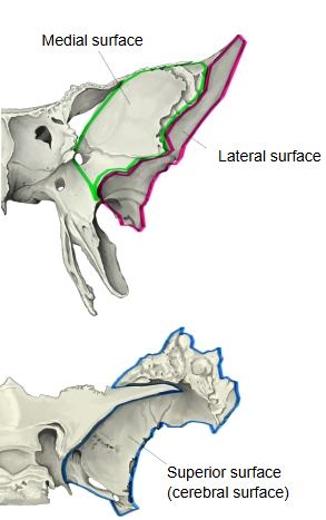
The features of each surface are summarized below.
| Surface | Features | |
|---|---|---|
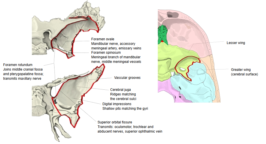
| 1 | Upper surface (cerebral surface) | · Concave and facing the brain; part of the middle cranial fossa. Features: digital impressions, cerebral juga, grooves for arteries and veins (meningeal vessels), and openings (foramen rotundum, foramen ovale, foramen spinosum). |
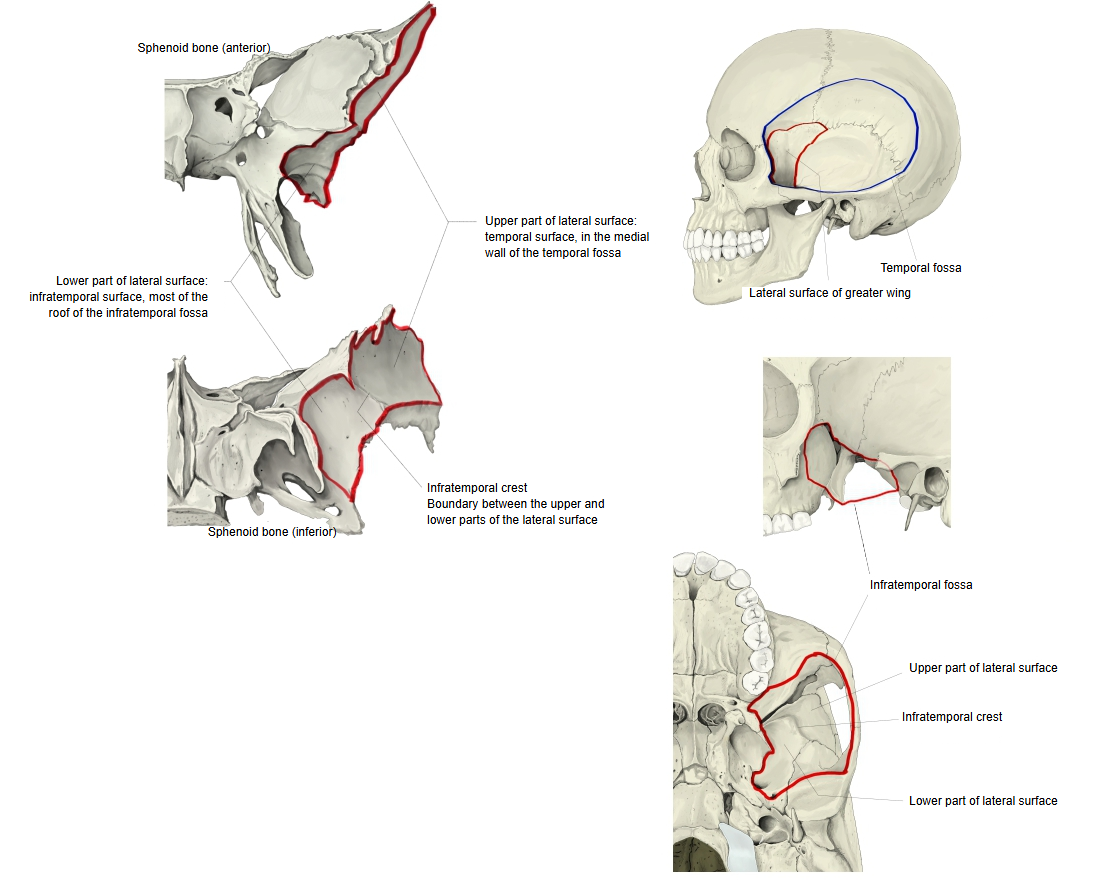
| 2 | Lateral surface | · In two parts, divided by the infratemporal crest: Upper part (temporal surface): part of the medial wall of the temporal fossa; part of the attachment of the temporalis. Lower part (infratemporal surface): most of the roof of the infratemporal fossa; part of the origin of the lateral pterygoid. Features: openings of the foramen ovale and foramen spinosum, the spine of the sphenoid, the infratemporal crest. |
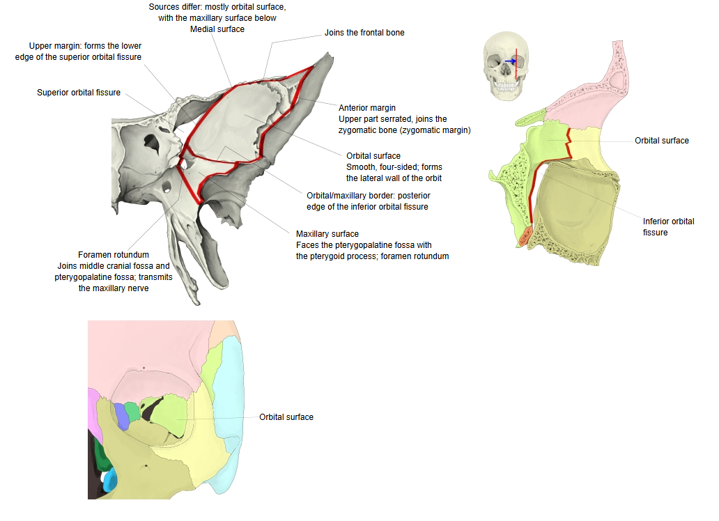
| 3 | Medial surface | · Its four-sided main part takes part in the lateral wall of the orbit, so it is called the orbital surface. · The small part below it is the maxillary surface; together with the front of the pterygoid process it faces the pterygopalatine fossa, and the foramen rotundum opens on it. · The border between the orbital and maxillary surfaces forms the back edge of the inferior orbital fissure. |
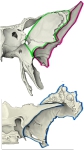 Surfaces of the greater wing
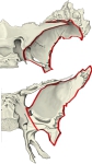 Upper (cerebral) surface
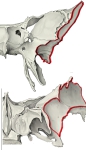 Lateral surface
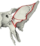 Medial (orbital and maxillary) surfaces
Margins
The features of each margin are summarized below.
| Margin | Features | |
|---|---|---|
| 1 | Upper margin | · Runs from the root of the greater wing up and out to its highest tip (= frontal margin). Medial part: sharp like a blade; forms the lower edge of the superior orbital fissure. Lateral part: a broad, rough surface, partly serrated and partly sharp like a chisel edge; joins the frontal bone in front and the parietal bone behind (its tip, the parietal margin, meets the parietal bone at the pterion). |
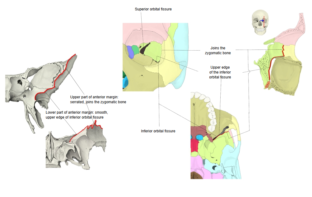
| 2 | Anterior margin | · Upper part: serrated; joins the zygomatic bone (= zygomatic margin). · Lower part: smooth; forms the upper edge of the inferior orbital fissure. |
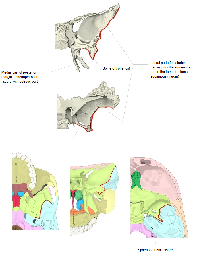
| 3 | Posterior margin | · Lateral (front) part: joins the squamous part of the temporal bone, so it is called the squamous margin. · Medial (back) part: forms the sphenopetrosal fissure with the petrous part of the temporal bone. Spine of the sphenoid: a process running downwards from where the two parts meet. |
- Margins of the greater wing
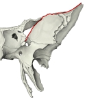 Upper margin
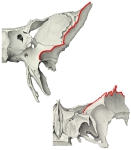 Anterior margin
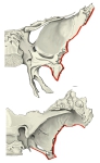 Posterior margin
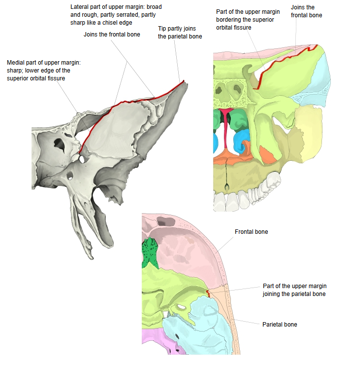
Openings
| Opening | Leads to / contents | |
|---|---|---|
| 1 | Foramen rotundum | From the middle cranial fossa to the pterygopalatine fossa; carries the maxillary nerve. |
| 2 | Foramen ovale | To the infratemporal fossa; carries the mandibular nerve, the accessory meningeal artery and emissary veins. |
| 3 | Foramen spinosum | Carries the middle meningeal artery and vein and the meningeal branch of the mandibular nerve. |
| 4 | Superior orbital fissure | Between the greater and lesser wings; carries the oculomotor, trochlear and abducent nerves, branches of the ophthalmic nerve and the superior ophthalmic vein. |
Reference websites
[Wikipedia]
· Greater wing of sphenoid bone, Wikipedia.
· Sphenoid bone, Wikipedia.
[Websites with illustrations]
· Bony landmarks of the sphenoid wings and pterygoid process (TeachMeAnatomy)
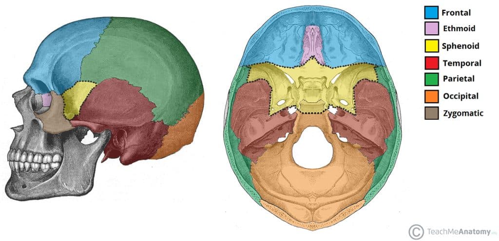
· Position of the sphenoid bone in the skull (TeachMeAnatomy)
· Greater wing of the sphenoid highlighted on the skull (Alamy)

Where it is found
Related terms
- Sphenoid bone
- Body of sphenoid
- Lesser wing
- Pterygoid process
- Foramen rotundum
- Foramen ovale
- Superior orbital fissure
References and sources
- Further reading: Greater wing of sphenoid bone, Wikipedia.
- Further reading: Sphenoid bone, Wikipedia.
- Federative International Programme for Anatomical Terminology (FIPAT). Terminologia Anatomica, 2nd ed. (TA2), 2019.