Neurocentral synchondrosis
· The term is not found in every anatomy textbook, but several sources describe and illustrate it.
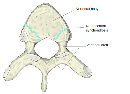
In other words, it is the paired cartilaginous union, one on each side, between the centrum and the neural arch of a vertebra that is still forming in a young child.
The names of the joints of the vertebrae are easily confused; they are summarised below.
the vertebrae
- Zygapophysial joint : (facet joint) between the superior and inferior articular processes of neighbouring vertebrae; a synovial joint (a joint in the narrow sense).
- Intervertebral symphysis : between the bodies of neighbouring vertebrae through the intervertebral disc (fibrocartilage); a cartilaginous joint.
- Neurocentral synchondrosis : in a young child, the hyaline cartilage joint between the centrum and the arch of the same vertebra; not a joint between two vertebrae.
A typical vertebra ossifies from three primary centres: one for the centrum and one for each half of the neural arch. The two neurocentral synchondroses join them, one on each side. They lie in the back and side parts of the future vertebral body, so in the adult the arch forms a small part of the body as well as the pedicles. They turn to bone in early childhood, so that the body and arch become one bone; the age varies with the region of the column, and sources differ. The neurocentral synchondrosis joins parts of a single vertebra and is not the joint between neighbouring vertebral bodies, which is the intervertebral symphysis. Before it closes it can look like a fracture line on X-ray or CT images. Compare the sphenooccipital synchondrosis of the skull and the epiphyseal cartilage of long bones, other cartilage plates that later ossify.
Reference sites
· Neurocentral synchondrosis, from Gray's Anatomy (figure)

Related terms
- Vertebra
- Intervertebral symphysis
- Cartilaginous joint
- Sphenooccipital synchondrosis
- Epiphyseal cartilage
- Vertebral body
- Vertebral canal
References and sources
- Further reading: Synchondrosis, Wikipedia.
- Further reading: Vertebra, Wikipedia.
- Federative International Programme for Anatomical Terminology (FIPAT). Terminologia Anatomica, 2nd ed. (TA2), 2019.