Vertebral canal (Latin: canalis vertebralis)
Also called the spinal canal
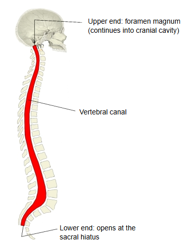
The space inside the vertebral column through which the spinal cord runs up and down.
More detail
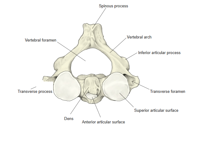
The vertebral canal is the space formed by the vertebral foramina of all the vertebrae lined up one above the other, joined by the intervertebral discs and ligaments. Its shape at each level follows the shape of the vertebral foramen, and it follows the curves of the vertebral column. Above, it opens into the cranial cavity through the foramen magnum; below, the sacral canal is its lowest part, which opens at the sacral hiatus. Its main features are:
| Part | Shape of the canal |
|---|---|
| Cervical | Widest; a broad triangle with blunt corners. |
| Thoracic | Narrow and round. |
| Lumbar | Wide and triangle-shaped with blunt corners. |
| Sacral canal | Becomes narrower as it goes down. |
Note: the vertebrae below are not drawn to the same scale.
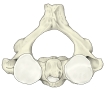 2nd cervical vertebra (axis)
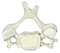 4th cervical vertebra
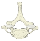 7th cervical vertebra
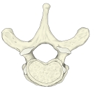 2nd thoracic vertebra
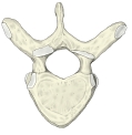 6th thoracic vertebra
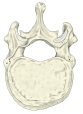 12th thoracic vertebra
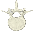 2nd lumbar vertebra
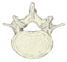 4th lumbar vertebra
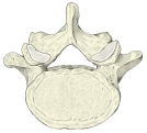 5th lumbar vertebra
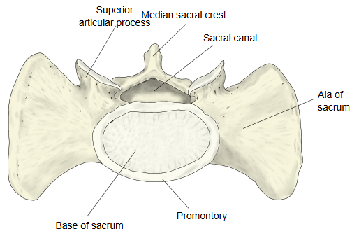 Sacrum (superior view)
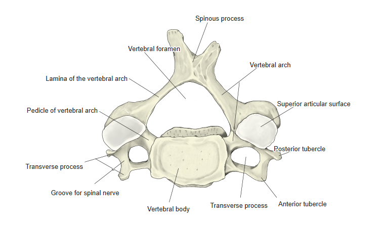
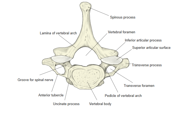
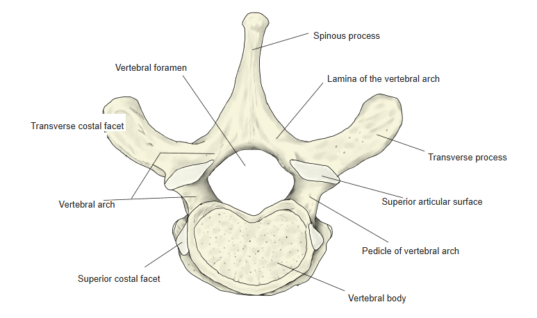
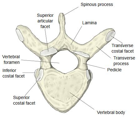
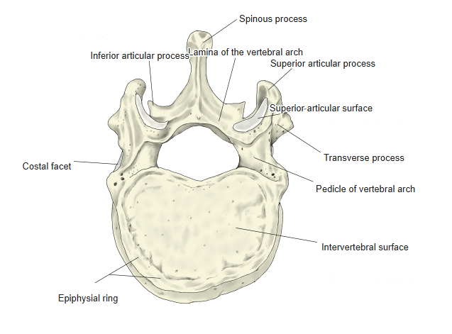
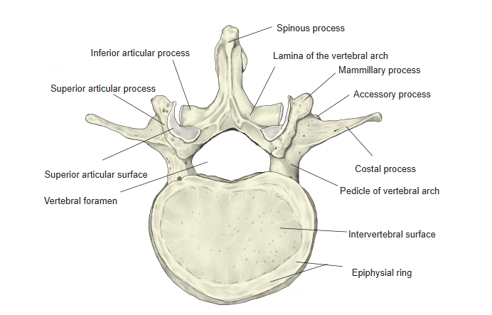
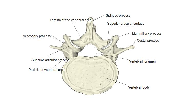
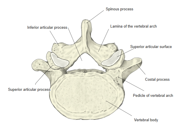
Its main contents are:
· The vertebral canal is the passage for the spinal cord (down to about the L1–L2 level) and, below it, the nerve roots of the cauda equina.
· The spinal cord is surrounded and protected by the meninges: dura mater, arachnoid mater and pia mater, with cerebrospinal fluid in the subarachnoid space.
· Blood vessels of the spinal cord: the anterior and posterior spinal arteries and the anterior and posterior spinal veins; the roots of the spinal nerves also run in the canal.
· Between the dura and the bone lies the epidural space, with fat and the internal vertebral venous plexus. Narrowing of the canal (spinal stenosis) can press on the spinal cord or the nerve roots.
Related terms
- Vertebral cavity
- Groove for spinal nerve
- Medullary cone
- Vertebral region
- Erector spinae
- Vertebral column
- Vertebral body
- Sacral canal
- Foramen magnum
- Sacral hiatus
- Spinal cord
- Dura mater
References and sources
- Further reading: Spinal cavity, Wikipedia.
- Further reading: Vertebral column, Wikipedia.
- Federative International Programme for Anatomical Terminology (FIPAT). Terminologia Anatomica, 2nd ed. (TA2), 2019.