Pterygoid tuberosity (Latin: tuberositas pterygoidea)
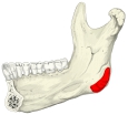 Pterygoid tuberosity
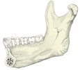 Mandible (medial surface)
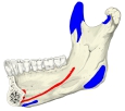 Muscle attachments (medial surface)
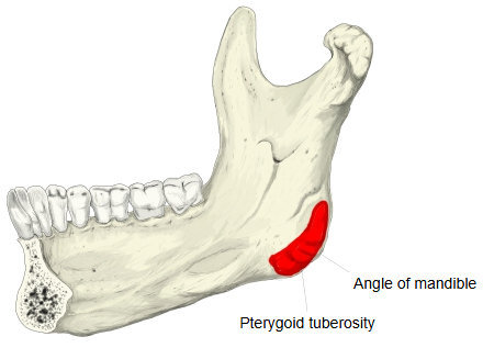
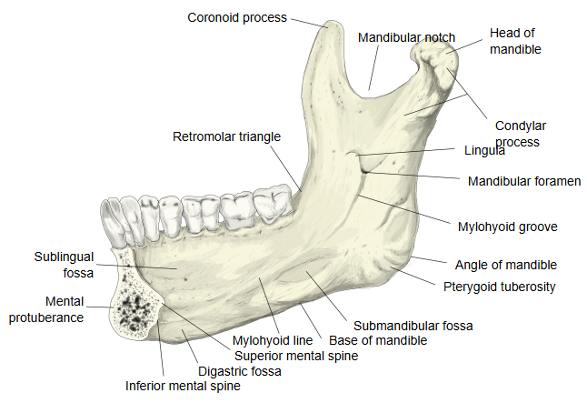
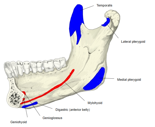
A roughened area on the inner surface of the mandible, just above and in front of the angle, where the fibres of the medial pterygoid are inserted. Its ridges are made by the tendinous fibres of the muscle, which runs down and back from the pterygoid fossa of the sphenoid bone. It lies below and behind the mandibular foramen; further forward, on the body of the mandible, runs the mylohyoid line.
· It is the inner counterpart of the masseteric tuberosity on the outer surface, where the masseter is inserted; together the two muscles form a sling around the angle.
Reference websites
[Wikipedia]
· Mandible, Wikipedia.
[Websites with illustrations]
· Mandible, inner surface, side view (Gray's Anatomy, fig. 177) (Wikimedia Commons)

· The mandible (TeachMeAnatomy)
· The mandible (Kenhub)
Related terms
- Pterygoid fossa
- Angle of mandible
- Ramus of mandible
- Masseteric tuberosity
- Masseter
- Mandible
- Mylohyoid line
- Mandibular foramen
References and sources
- Further reading: Mandible, Wikipedia.
- Federative International Programme for Anatomical Terminology (FIPAT). Terminologia Anatomica, 2nd ed. (TA2), 2019.