Masseteric tuberosity (Latin: tuberositas masseterica)
· The masseter inserts not only on the masseteric tuberosity but also on the rest of the lateral surface of the ramus of the mandible, up to the coronoid process.
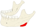 Masseteric tuberosity
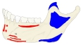 Muscle attachments (lateral surface)
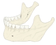 Mandible (lateral)
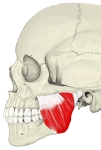 Masseter
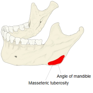
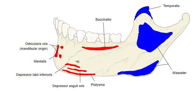
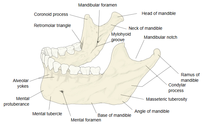
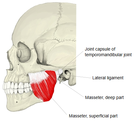
Anatomy textbooks describe it as follows.
The outer surface of the ramus of the mandible is mostly smooth, but near the angle of the mandible, on its lower part just above and in front of the angle, lies the large masseteric tuberosity, where the masseter inserts. It is often marked by oblique ridges, which become more prominent where the masseter is strong.
Other English descriptions put it the same way.
It is a rough area on the outer surface of the angle of the mandible for the attachment of the masseter. On the inner surface of the angle lies the matching pterygoid tuberosity, for the medial pterygoid.
Reference websites
[Wikipedia]
· Mandible, Wikipedia.
· Masseter muscle, Wikipedia.
[Websites with illustrations]
· The mandible: structure and attachments (TeachMeAnatomy)
· Masseter muscle: origin, insertion, function (Kenhub)
Related terms
- Mandible
- Masseter
- Angle of mandible
- Ramus of mandible
- Base of mandible
- Zygomatic arch
- Temporomandibular joint
References and sources
- Further reading: Mandible, Wikipedia.
- Federative International Programme for Anatomical Terminology (FIPAT). Terminologia Anatomica, 2nd ed. (TA2), 2019.