High endothelial venule
Also called HEV
Overview
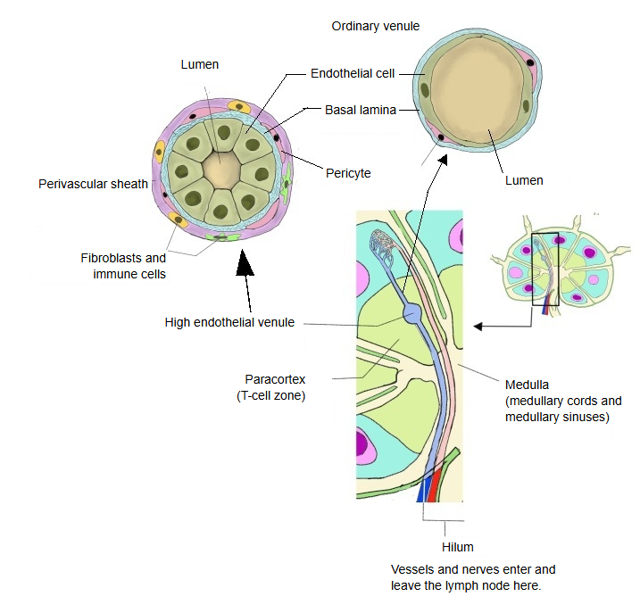
Structure of the wall
The table summarises the layers of the HEV wall (simplified).
| Layer | Components | Notes | |
|---|---|---|---|
| 1 | Endothelium | Tall cuboidal to columnar endothelial cells | Unlike the flat endothelium of an ordinary venule. Helps lymphocytes pass from the blood stream into the tissue. |
| 2 | Basement membrane | Basal lamina (type IV collagen and other proteins) | Supports the endothelial cells; in HEVs it may be somewhat discontinuous, which can make it easier for lymphocytes to pass. |
| 3 | Pericytes | Supporting cells attached around the vessel | Take part in stabilising the vessel and in regulating blood flow and permeability; in HEVs they may be fewer than around ordinary capillaries. |
| 4 | Perivascular sheath | Connective tissue, fibroblasts, a few immune cells | Supports the HEV and forms the small space where lymphocytes gather; a thin sheath rather than a thick layer. |
Function
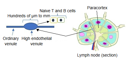
· They are the route by which naive T cells and naive B cells pass from the blood stream into the lymph node.
· The surface of the endothelial cells carries molecules that bind selectins (special sugar-bearing molecules, the peripheral node addressin, that bind L-selectin on lymphocytes), so that passing lymphocytes are selectively captured and roll along the wall. Chemokines on the endothelium then activate integrins on the lymphocytes, which stick firmly and squeeze between the endothelial cells into the tissue.
Development
When HEVs form
1. Fetal period: formation of the lymph nodes (before HEVs)
· When the rudiments of the lymph nodes (lymph node anlagen) appear, their vessels are at first lined by ordinary flat endothelial cells.
· At this stage there are no mature HEVs yet.
· Because HEVs are special vessels for letting lymphocytes in, they develop only when the surroundings in which lymphocytes work are ready.
2. After birth and in infancy: HEVs begin to form
· After birth, as lymphocytes begin to gather around them, the endothelium starts to express HEV markers such as the L-selectin ligand (peripheral node addressin, PNAd).
· At the same time the endothelial cells change to a cuboidal or columnar shape and become "high".
So the formation of HEVs needs an interaction (crosstalk) with lymphocytes.
3. Childhood: functional maturation of HEVs
· As the distribution of T and B cells within the lymph nodes becomes stable during childhood, the HEVs mature as well.
· From then on, lymphocyte homing through L-selectin and PNAd works fully.
In the adult, HEVs are kept up by lymphocytes
HEVs disappear if lymphocytes stop flowing into them.
· In experiments, when the entry of lymph or lymphocytes is cut off, HEVs flatten into ordinary-looking venules.
· When lymphocytes are supplied again, the endothelium becomes high again.
⇒ The HEV is not a fixed structure but a dynamic one, kept up by the lymphocytes. (Much of this is known from animal studies.)
HEV-like vessels in inflammation (adults)
· In tissues with strong, long-lasting inflammation (for example chronic inflammation or autoimmune disease), vessels like HEVs can be induced for a time outside the lymph nodes.
· These are called "HEV-like vessels"; they differ somewhat from ordinary HEVs but work in a similar way.
Summary
· Mature HEVs are not present in the fetal lymph node rudiments.
· HEVs begin to form after birth, when lymphocytes start to flow in.
· They mature during childhood.
· In the adult they are kept up by the continuing inflow of lymphocytes; when the inflow stops, they disappear.
Reference websites
[Wikipedia]
· High endothelial venules, Wikipedia.
· Lymph node, Wikipedia.
Related terms
- Lymph node
- B lymphocyte
- Efferent lymphatic vessel
- Hilum of lymph node
- Diffuse lymphoid tissue
- Lymphoid nodule
- Aggregated lymphoid nodules
References and sources
- Further reading: High endothelial venules, Wikipedia.
- Further reading: Lymph node, Wikipedia.
- Federative International Programme for Anatomical Terminology (FIPAT). Terminologia Anatomica, 2nd ed. (TA2), 2019.