Basement membrane (Latin: membrana basalis)
- Overview
- Structure
- Where it is found
- Reference websites
- (Wikipedia, websites with illustrations)
- Related terms
Overview
· Its thickness is roughly 50–150 nm in most places (well under a micrometre), so it is best seen with the electron microscope; with special stains it shows as a fine line under the epithelium. Some basement membranes, such as the glomerular basement membrane, are much thicker.
It also acts as a barrier: an early cancer that has not yet broken through the basement membrane is called carcinoma in situ.
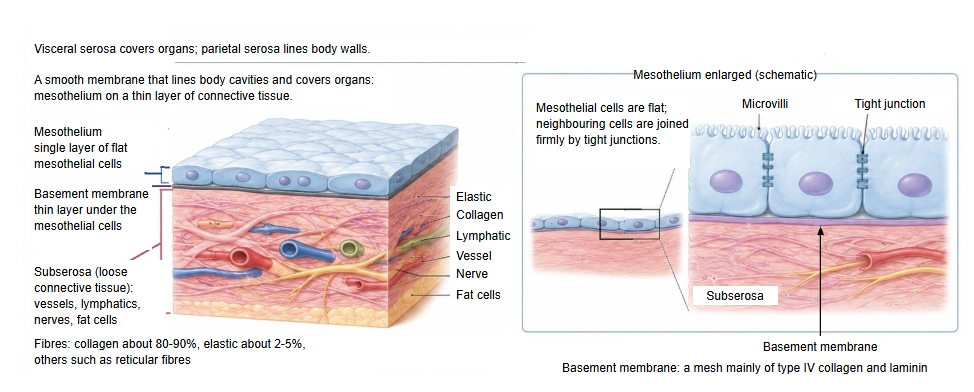
The figure below shows the basement membrane in a serous membrane, under the mesothelium.
Structure
The basement membrane has the following three layers.
of the
basement
membrane
- Lamina lucida : lies just under the epithelial cells; the adhesion molecule laminin, which is abundant here, fixes the cells to the basement membrane (through integrins of the hemidesmosomes).
- Lamina densa : a strong, net-like filter of type IV collagen, coated with the proteoglycan perlecan; nidogen (entactin) links laminin to it.
- Reticular lamina : fibres of type III collagen (reticular fibres) made by the connective tissue, tied to the lamina densa by anchoring fibrils of type VII collagen; the base that fixes the lamina densa to the connective tissue beneath.
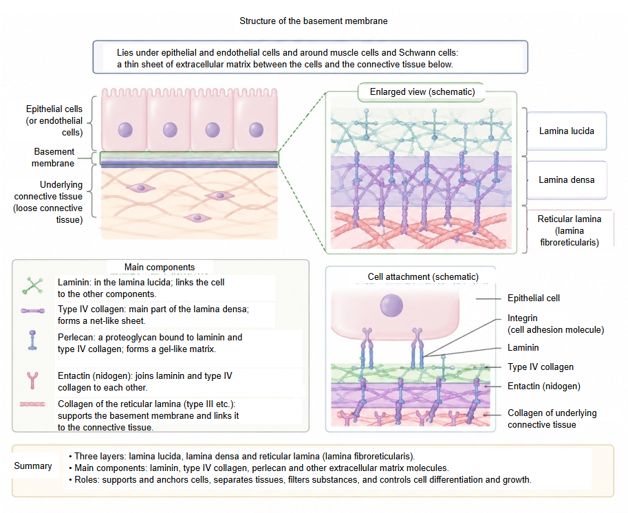
The lamina lucida and the lamina densa together form the basal lamina, made by the epithelial cells. Sources differ in naming: some use "basement membrane" and "basal lamina" as the same thing; others keep "basement membrane" for the basal lamina plus the reticular lamina.
Where it is found
Basement membranes are found mainly in the following places.
| Group | Where exactly | Main role there | |
|---|---|---|---|
| 1 | Body surface and linings of hollow organs (under epithelial cells) | Skin (boundary between the epidermis and the dermis) | Anchors the epidermis firmly to the dermis and protects it against outside forces. |
| Digestive tract (oesophagus, stomach, intestine, etc.) | Controls the absorption of nutrients and helps to keep out pathogens. | ||
| Respiratory tract (trachea, bronchi, etc.; under a pseudostratified epithelium) | The base for the ciliated cells and other cells that sweep out inhaled particles. | ||
| 2 | Coverings of organs and body cavities (under mesothelial cells) | Serous membranes (pleura, peritoneum, pericardium) | The base of the mesothelium, which reduces friction between organs; controls the movement of fluid. |
| 3 | Blood and lymphatic vessels (under endothelial cells) | Capillaries, arterioles, etc. | Keeps the vessel wall in shape and stops blood components leaking out. (Lymphatic capillaries have little or none.) |
| 4 | Special organs and filter membranes | Glomerulus of the kidney (glomerular basement membrane) | Filters the blood to make urine; the thickest basement membrane in the body. |
| Pulmonary alveoli (alveolar septum) | Extremely thin (the basement membranes of the alveolar cells and of the capillary are fused) so that gas exchange is easy. | ||
| 5 | Sheaths around single cells (often called the external lamina) | Skeletal and cardiac muscle fibres (also smooth muscle cells) | Protects the cell membrane from damage by strong muscle contraction (a cushion). |
| Peripheral nerves (Schwann cells) | Wraps the nerve fibres and helps to protect and regenerate the nerve. | ||
| Fat cells | Wraps each fat cell completely and keeps its shape. |
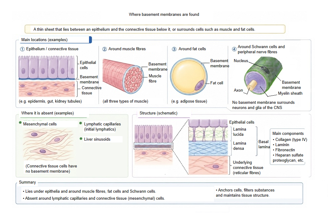
It is absent or incomplete around lymphatic capillaries, liver sinusoids and connective tissue cells, and there is none around the neurons and glia inside the central nervous system.
Reference websites
[Wikipedia]
· Basement membrane, Wikipedia.
· Basal lamina, Wikipedia.
[Websites with illustrations]
· Extracellular matrix and basement membrane (diagram) (Wikimedia Commons)

· Oral mucosa with the basement membrane under the epithelium (Wikimedia Commons)

· Basement membrane: anatomy and function (Kenhub)
Related terms
- Smooth muscle
- Alveolar septum
- Pseudostratified epithelium
- Glomerular basement membrane
- Serous membrane
- Mesothelium
- Epidermis
- Dermis
- Alveolar cells
References and sources
- Further reading: Basement membrane, Wikipedia.
- Further reading: Basal lamina, Wikipedia.
- Federative International Programme for Anatomical Terminology (FIPAT). Terminologia Anatomica, 2nd ed. (TA2), 2019.